Wednesday, December 9, 2015

Not allowing kids to play football is "our moral duty": Bennet Omalu, MD

In a recent New York Times opinion piece, pathologist Bennet Omalu strongly endorsed a policy that children should not play football. Here are excerpts from his essay:

"We've known since 1964 that cigarette smoking is harmful to your health. We’ve known for more than 40 years that alcohol damages the developing brain of a child. We’ve known since the mid-70s that asbestos causes cancer and other serious diseases. Knowing what we know now, we do not smoke in enclosed public spaces like airplanes; we have passed laws to keep children from smoking or drinking alcohol; and we do not use asbestos as an industrial product.

"If a child who plays football is subjected to advanced radiological and neurocognitive studies during the season and several months after the season, there can be evidence of brain damage at the cellular level of brain functioning, even if there were no documented concussions or reported symptoms. If that child continues to play over many seasons, these cellular injuries accumulate to cause irreversible brain damage, which we know now by the name Chronic Traumatic Encephalopathy, or C.T.E., a disease that I first diagnosed in 2002."

It should be noted that the CTE brain bank at Boston University found evidence of CTE in the brains of 131 out of 165 athletes who played professional, college, or high school football. In other words, based on this sample size, the risk of getting CTE from playing football for a relatively extended period is higher than the risk of getting lung cancer from smoking.

Since we have a legal age at which individuals are allowed to smoke, drink alcohol, and drive, it stands to reason that engaging in an activity that is likely to cause brain damage should also only be engaged in by individuals old enough to make an informed decision, says Omalu. 

Thanks to Dr. Chad Vanderbilt for alerting me to this article.

Tuesday, November 24, 2015

The Mercado Brain Cutting Device launched at the University of Colorado

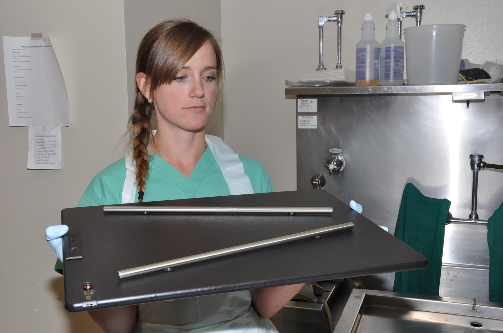
Thanks to a hand-made gift from University of Alabama neuropathology fellow Juan Mercado, MD, our residents on autopsy rotation this month had the opportunity yesterday to inaugurate the use of The Mercado Brain Cutting Device (MBCD). Made from tools easily found at any hardware store, the device allows prosectors to make reliably even 1-cm thick coronal brain slices for optimal demonstration of gross anatomy and pathology. University of Colorado pathology residents Abby Richmond, MD (PGY-III) and Sammie Roberts, MD (PGY-I) used the device to great advantage as demonstrated by the exquisitely presented brain slices laid out for inspection.

Dr. Sammie Roberts with the MBCD 
Dr. Abby Richmond makes the first cut

Sections are even and uniform in thickness

The finished product

Drs. Moore, Richmond, and Roberts (left to right) examining the coronal sections

Much appreciation to Dr. Mercado for gifting this device, which he describes as a "limited edition (1 or 1)", to our department. We will undoubtedly have more meticulous brain cutting sessions henceforth thanks to Dr. Mercado's efforts.

Wednesday, November 18, 2015

A 60-year-old woman with a left occipitotemporal brain lesion

We were sent this case in consultation to rule out infiltrating neoplasm. The diagnosis is amyloid beta related angiitis (ABRA). In this case, no infarction was present in the specimen, but white matter rarefaction (the presumed pathologic correlate to the leukoaraiosis reported on imaging) is present.

Granulomatous vasculitis associated with pink hyaline material in vessel walls

The pink hyaline material is shown to be beta-amyloid by immunohistochemistry

White matter rarefaction (upper half of picture) correlating with leukoaraiosis seen on imaging

Tuesday, November 17, 2015

Best Post of June 2015: First Day at 2015 American Association of Neuropathologists Meeting - The Special Course

The next in our "Best of the Month" series comes from 11 June 2015, where we harken back to the first day at the American Association of Neuropathologists (AANP) annual meeting held in my newly adopted home of Denver, Colorado:


The "Special Course" which typically launches the annual AANP meeting was anything but typical this year. Rather than the usual series of research presentations which has characterized the first day of the meeting, the 2015 edition of the "Special Course" focused on need-to-know practical topics. Dr. Beatriz Lopes put together a program that offered something for everyone -- from trainee to seasoned practitioner. The morning started with a presentation by Dr. Caterina Giannini on primary CNS lymphoma, as well as its mimickers and precursors. Far from being a straightforward diagnosis, lymphoma -- particularly in the setting of corticosteroid therapy -- can be mistaken for anything from multiple sclerosis to infarct. Next, two eminent scholars from Paris, Drs. Francoise Gray and Elisabeth Tournier-Lasserve, presented a three-part lecture on the hereditary non-amyloid small vessel diseases of the brain. Then Charles Eberhart showed up from Johns Hopkins to provide a primer on ophthalmic pathology, providing a practical approach to eye specimens. For example, Dr. Eberhart discussed the significance of uveal granulomatosis in orbital exenteration specimens. The presence of this finding should be noted in the pathology report as it portends an increased risk for sympathetic ophthalmia. Dr. Arie Perry appeared next with a presentation on the molecular characterization of brain tumors using immunohistochemical surrogates -- an approach particularly appreciated by neuropathologists who work at smaller institutions which may not have the resources to perform sequencing and other more advanced laboratory tests. After lunch, the focus was redirected to neuropathology training. Drs. Marc Del Bigio and Suzanne Powell discussed the state of neuropathology training abroad and in the United States, respectively. Finally, a lively discussion was initiated by a panel featuring Drs. Jeff Golden, Dennis Dickson, Liz Cochran, and myself organized under the title "The Professional Market for Neuropathology Trainees". Several audience members talked about their view of the profession and how its many facets are reflected in the training we give fellows and how that training impacts the preparedness of trainees for the job market. Overall, it was engaging day which, as audience member John Donahue put it, was "well worth the price of admission!"

Tuesday, November 10, 2015

Best Post of May 2015: "Bad Ass" CrossFitter and Neuropathologist Greg Fuller set to compete in kettlebell competition for brain cancer research

The next in our "Best of the Month" Series comes from Wednesday, May 20, 2015:


Elite Neuropathologist
Elite Athlete
It's no secret that Dr. Greg Fuller (pictured) is the fittest neuropathologist in the world. What his colleagues might not know is that he is the 67th fittest man in the world for his age class (60+). Greg's wife, Tina, describes him as a "bad ass CrossFitter". As a former owner of a CrossFit "box" myself, I know what it takes to achieve Greg's level in the Sport of Fitness. It requires true grit. There are many of us who would also consider Greg a "bad-ass neuropathologist" -- in the very best sense of that phrase. He's bringing together his love of fitness with his commitment to the study of CNS malignancies by participating in the Kettlebells For Brain Cells competition in Boerne, Texas on June 13. (So, he has a legitimate reason for not showing up at the concurrent AANP annual meeting. in Denver.) All proceeds from the competition will be going to the American Brain Tumor Association. The event is being organized by a brain tumor patient at MD Anderson Cancer Center, where Greg is Chief Neuropathologist. This is a shining example of a neuropathologist reaching out to a patient in a very personal way. Best of luck in the competition, Greg. Crush it!

Wednesday, October 28, 2015

Best Post of April 2015 - The Tumor Biomarker Series: BRAF

The next in our "Best of the Month" series comes from Tuesday, April 28, 2015:


BRAF gene (v-Raf murine sarcoma viral oncogene homolog B)

BRAF+ IHC (correlates with V600E mutation) in ganglioglioma
Aberrant constitutive activation of BRAF tends to be seen in cerebellar and midline pilocytic astrocyomas whereas the activating point mutation at BRAF V600E is more likely to be seen in cerebral examples. The V600E point mutation is also observed in other low-grade gliomas and glioneuronal neoplasms, including approximately two-thirds of pleomorphic xanthoastrocytomas, and lower percentages of ganglioglioma, desmoplastic infantile ganglioglioma, dysembrioplastic neuroepithelial tumor, and papillary craniopharyngioma. Although less common, diffusely infiltrative gliomas including glioblastoma, particularly the epithelioid variant, may also demonstrate the V600E point mutation -- making this biomarker potentially less useful as a diagnostic tool in distinguishing low-grade gliomas from high-grade ones.

Friday, October 23, 2015

Juvenile psammomatoid ossifying fibroma in 22-year-old male with eyelid droop

This lesion was arising from the nasal sinuses but was growing into the anterior skull base and pushing into the left orbitofrontal region. At first low-power glance, I thought it was a bread-and-butter psammomatous meningioma. But it revealed itself when targeted with a 20X objective lens:


Thursday, October 22, 2015

Brain Tumor Rhapsody by Dr. Arie Perry

Arie Perry, MD
I have written about Dr. Arie Perry's incredible musical talent before, and how he has applied it to neuropathology education. Well, he's brought that talent to a whole new level through a collaboration with the San Francisco Bay Area's vocal ensemble Musaic.  With Virchow as his muse, Arie outlines major biomarkers in the diagnosis of CNS tumors. This magnum opus is called Brain Tumor Rhapsody, and its Dr. Perry's first-ever educational music video.This is an epic musical and neuropathological achievement! Many thanks to Dr. Gabrielle Yeaney of the Cleveland Clinic for alerting me to this remarkable video. Check it out on YouTube!


Gabrielle A. Yeaney, MD

Thursday, October 15, 2015

Heterotopic neuron in a patient with epilepsy

The patient is a 22-year-old female with intractable epilepsy who underwent resection of an epileptogenic region of the left lateral temporal lobe. In addition to mild cortical dyslamination (not depicted) and Chaslin's subpial gliosis (pinkish band on top surface of brain in photomicrograph), there were an increased number of individual heterotopic neurons within the neocortical molecular layer. The heterotopic neuron pictured below is also disoriented, with its axon projecting tangentially rather than perpendicularly to the the pial surface.


Monday, October 12, 2015

Best Post of March 2015: The asinine reason why the name JCD was converted to CJD

The next in our "Best of the Month" series is from March 25, 2015.


Originally referred to as Jakob-Creutzfeldt disease (and believed by many to rightfully be called simply Jakob's disease), we now refer to this prion disease as Creutzfeldt-Jakob disease. Why? It all stems from a Dr. C. Joseph Gibbs, a colleague of D. Carleton Gajdusek's at the National Institutes of Health, who worked on this disease back in the 1960's. Here's the explanation, lifted from Nobel laureate Stanley Prusiner's Madness and Memory: The Discovery of Prions - A New Biological Principle of Disease:


"Alfons Jakob wrote a paper in 1921 describing several cases of progressive dementia with widespread neuronal loss in the brain. A year earlier, Hans Creutzfeldt had described the brain of a woman who died after a prolonged series of seizures. He found widespread vacuolation, but some medical scientists believe that his patient died of a seizure disorder complicated by hypoxic brain damage and doubt that she had what became known as CJD. So why did the named Jakob and Creutzfeldt become flipped in an important report on the disease, regarding the transmission to a chimpanzee? Twenty years passed before I found the answer, in conversation with Gibbs. 'When I was writing my first paper on the transmission of Jakob-Creutzfeldt disease to an ape,' he told me, 'I wanted to rename the disease Gibbs disease. I didn't think this would be acceptable to the scientific and medical communities, so I decided to reverse the names, because my first name is Clarence and my middle name is Joseph and my initials are C.J.' Thus did the diease become known as Creutzfeldt-Jakob disease, or CJD for short - a case of mind-boggling scientific mischief." 

Gibbs (left) and Gajdusek with a New Guinea kuru patient in 1972.
 
A remarkable case ofhubris, although from what I hear about Prusiner, he is not one to point fingers at those who grasp credit for the work of others.Gajdusek, also a Nobel prize winner, once wrote inhis diary the following about Prusiner:


""I never heard a word of original thought from you nor read such ideas in anything you authored for which I did not recognize immediately its source, which you always went out of your way to obscure. You a heretic? You a martyr? You a defender of unacceptable ideas? Bullshit! You shrewdly jumped onto a bandwagon of creative ideas and experimental work and shrewdly got on to the winning cart, proclaiming outrageously in press and media it was yours! I respect you less and less as your despicable game succeeds and you bask in your coveted fame."

But then again, Gajdusek himself, who died in 2008 at age 85, was no paragon of virtue.In the course of his research trips in the South Pacific to study kuru , Gajdusek had brought 56 mostly male children back to live with him in the United States, and provided them with the opportunity to receive high school and college education. He was later accused by one of these, now an adult man, of molesting him as a child. In fact, seven men testified in confidentiality about Gajdusek having had sex with them when they were boys.  Gajdusek was charged with child molestation in April 1996, based on incriminating entries in his personal diary and statements from a victim. He pleaded guilty in 1997 and, under a plea bargain, was sentenced to 12 months in jail. After his release in 1998, he was permitted to serve his five-year unsupervised probation in Europe. He never returned to the United States and ultimately died in Norway.

Neuropathology Blog is Signing Off

Neuropathology Blog has run its course. It's been a fantastic experience authoring this blog over many years. The blog has been a source...