Showing posts with label history. Show all posts

Friday, August 10, 2018

Best Post of June 2018: Who was Rosenthal?

Of the ten posts from June 2018, my favorite was "Who was Rosenthal?" from June 22. So, here's the next in our "Best of the Month" series:

A Rosenthal fiber in pilocytic astrocytoma

"In 1898, the German pathologist Werner Rosenthal noted elongated inclusions within the gliotic edge of a syringeal cavity of an ependymoma. Assigned to write the case report by a senior mentor while serving as a “first assistant” at the University of Erlangen, Rosenthal colorfully described these inclusions as a “glossy formation of little bulbs or wavy sausages with one thick and one pointed end.”….  His supposition that they were related to glial fibers would prove surprisingly insightful. Not until some 20 years later did Bielschowsky and Unger use the term 'Rosenthal fibers' when describing structures in the gliotic capsule of a cystic teratoid tumor."

Quoted from:  F.J. Wippold, A. Perry and J. Lennerz. Neuropathology for the Neuroradiologist: Rosenthal Fibers. American Journal of Neuroradiology May 2006, 27 (5) 958-961.

Thursday, July 5, 2018

More on Rosenthal

Thanks to Dr. Gregg B. Wells from Texas A&M University for directing me to an article authored by Dr. Hans Goebel of Charite-Universitätsmedizin in Berlin regarding the life and accomplishments of neuropathologist Werner Rosenthal (1870-1942). Dr. Wells was prompted to write to me by my recent post entitled "Who was Rosenthal?". For those who are interested in Dr. Rosethal's travails as a Jew in Nazi Germany forced into exile in India, I encourage you to read Dr. Goebel's open-access article.

Dr. Werner Rosenthal with his wife and daughter in 1917.
(Taken from Dr. Goebel's article in Clinical Neuropathology)

Wednesday, April 11, 2018

There is a neuropathology museum in Lima, Peru

The Institute of Neurological Science (INCN) in Lima  houses the only "Brain Museum" in Latin America. The  collection contains over 3000 samples from which around 300 are on permanent display, including brains with diverse cerebral diseases and anomalies as well as fetal brains with abnormalities.  Unique photomicrographs and other items are also on display.

Wednesday, March 14, 2018

On Einstein's Birthday, We Take a Second Look at His Brain

On this date 139 years ago, Albert Einstein was born in Ulm, Germany. We take this occasion to republish a post from November 21, 2012 entitled: Photos reveal unique features of Einstein's cerebral cortex:

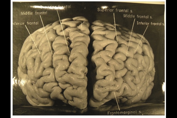
Photographs taken shortly after his death, but never before analysed in detail, have now revealed that Einstein’s brain had several unusual features, providing clues about the neural basis of his extraordinary mental abilities.


Nature.com reports that, while doing Einstein's autopsy, the pathologist Thomas Harvey removed the physicist's brain and preserved it in formalin. He then took dozens of black and white photographs of it before it was cut up into 240 blocks. Now, anthropologist Dean Falk of Florida State University in Tallahassee and her colleagues have obtained 12 of Harvey’s original photographs from the National Museum of Health and Medicine in Silver Spring, Maryland, analysed them and compared the patterns of convoluted ridges and furrows with those of 85 brains described in other studies.Many of the photographs were taken from unusual angles, and show structures that were not visible in photographs that have been analysed previously. The analysis was recently published today in the journal Brain. The most striking observation, says Falk, was “the complexity and pattern of convolutions on certain parts of Einstein's cerebral cortex”, especially in the prefrontal cortex, and also parietal lobes and visual cortex.

The autopsy revealed that Einstein’s brain was smaller than average and subsequent analyses showed all the changes that normally occur with ageing. Nothing more was analysed, however. Harvey stored the brain fragments in a formalin-filled jar in a cider box kept under a beer cooler in his office. Decades later, several researchers asked Harvey for some samples, and noticed some unusual features when analysing them.
A study done in 1985 showed that two parts of his brain contained an unusually large number of non-neuronal cells called glia for every neuron2. And one published more than a decade later showed that the parietal lobe lacks a furrow and a structure called the operculum3. The missing furrow may have enhanced the connections in this region, which is thought to be involved in visuo-spatial functions and mathematical skills.
AFP/Getty Images
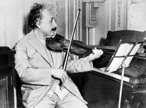
Einstein was a keen violinist, which may account for an overdeveloped section of his brain that deals with the left hand.
The prefrontal cortex is important for the kind of abstract thinking that Einstein would have needed for his famous thought experiments on the nature of space and time, such as imagining riding alongside a beam of light. The unusually complex pattern of convolutions there probably gave the region and unusually large surface area, which may have contributed to his remarkable abilities.
Falk and her colleagues also noticed an unusual feature in the right somatosensory cortex, which receives sensory information from the body. In this part of Einstein’s brain, the region corresponding to the left hand is expanded, and the researchers suggest that this may have contributed to his accomplished violin playing.
According to Sandra Witelson, a behavioural neuroscientist at McMaster University in Hamilton, Canada, who discovered that the parietal operculum is missing from Einstein’s brain, the study’s biggest contribution may be in encouraging further studies. “It makes clear the location and accessibility of photographs and slides of Einstein's brain,” she says. “This may serve as an incentive for other investigations of Einstein's brain, and ultimately of any consequences of its anatomical variations.”

Wednesday, March 7, 2018

The meticulously extracted nervous system of a 19th-century woman on display at Hahnemann Medical College

Last summer I put up a post about a remarkable whole nervous system dissection that was carried out at the University of Colorado School of Medicine. The inimitable Dr. Mark Cohen recently sent me an article about a similar dissection performed at Hahnemann Medical College in Philadelphia by Dr. Rufus B. Weaver. The dissection, which took place in 1888 over the course of five months, was performed on a 35-year-old woman who had given permission for her body to be used for the furtherance of science.

Dr. Rufus B. Weaver and the nervous system of Harriet Cole

An excerpt from the article appearing in Atlas Obscura:
According to the History of the Homoeopathic Medical College of Pennsylvania, Dr. Weaver told a fellow doctor about Harriet during a trip to the U.K., after his extraction of the nervous system. He didn’t mention the completion of the dissection. The doctor’s response: “It is impossible, there is no such thing in all this United Kingdom, and if it had been possible it would have been done by some one.” Dr. Weaver replied quietly: “So it has, by some one in the States.” 
In an article for Homeopathic World in August 1892, Dr. Alfred Heath was far more generous about Dr. Weaver’s accomplishment. He called it “a marvel of patience and skill in dissection, the likes of which has never been seen before.”

A dissection similar to that of Dr. Weaver's done at the University of Colorado in 2017 by Shannon Curran

Thursday, June 1, 2017

Best Post of February 2017: Hunched Over a Microscope, Santiago Ramon y Cajal Sketched the Secrets of How the Brain Works

The next in our "Best of the Month" series comes from February 20, 2017:


Last month, the Weisman Art Museum in Minneapolis opened a traveling exhibit that is the first dedicated solely to Ramón y Cajal’s work. According to a New York Times article, it will make stops in Minneapolis; Vancouver, British Columbia; New York City; Cambridge, Mass.; and Chapel Hill, N.C., through April 2019.

Ramon y Cajal in his laboratory, circa 1885
 Thanks to Drs. Mark Cohen and John Evans for alerting me to this exhibit.

Friday, March 3, 2017

Why is the confluence of the cerebral venous sinuses called the "torcula"?

Torcula is derived from a Latin word meaning to “twist” and was also used to refer to a wine press. Within the cranium the venous sinuses come together at the back of the skull in a structure called the confluence of the sinuses. This cavity has four large veins radiating from it, supposedly resembling the spigots that pour dark purple juice out of the four sides of the ancient wine press used to squeeze grapes with a handled screw on the top. The same stem is found in common words such as torture and tortuous.

Source: https://www.dmu.edu/dose/2009/11/anatomy-word-of-the-month-torcular-herophili/

Monday, February 20, 2017

Hunched Over a Microscope, Santiago Ramon y Cajal Sketched the Secrets of How the Brain Works


Last month, the Weisman Art Museum in Minneapolis opened a traveling exhibit that is the first dedicated solely to Ramón y Cajal’s work. According to a New York Times article, it will make stops in Minneapolis; Vancouver, British Columbia; New York City; Cambridge, Mass.; and Chapel Hill, N.C., through April 2019.

Ramon y Cajal in his laboratory, circa 1885
 Thanks to Drs. Mark Cohen and John Evans for alerting me to this exhibit.

Wednesday, December 7, 2016

Neuropathology History: Désiré Magloire Bourneville (1840-1909)

Désiré Magloire Bourneville (1840-1909)

Born in Garenciéres, France. From 1905 until his death, Bourneville headed the Foundation Vallée for the Study of Feebleminded Children.  "Was recognized as the leading continental authority on all aspects of mental abnormality of children. Most of his neuropathologic work was on idiocy. His description of tuberous sclerosis, since known as Bourneville's disease, appeared in 1880."

Source: Haymaker, Webb (Army Institute of Pathology). Guide to the exhibit on the history of neuropathology. Presented at the annual meetings of the American Psychiatric Association (Washington, DC, May 17-20, 1948) and the American Neurological Association (Atlantic City, June 14-17, 1948).

Tuesday, December 6, 2016

Neuropathology History: Sir Edward Farquhar Buzzard (1871-1945)

"Born in London... Became Regius Professor of Medicine at Oxford University, and consulting physician at St. Thomas's Hospital (1928). In the field of neuropathology, he is well known for his textbook in collaboration with Greenfield (1921)... Important also were his studies on myasthenia gravis (to which he contributed the term 'lymphorrhages') (1905), chronic progressive cerebral softening (1906),... delayed traumatic apoplexy (1909), and epidemic encephalitis (1919)."

Sir Edward Farquhar Buzzard (1871-1945)

Source: Haymaker, Webb (Army Institute of Pathology). Guide to the exhibit on the history of neuropathology. Presented at the annual meetings of the American Psychiatric Association (Washington, DC, May 17-20, 1948) and the American Neurological Association (Atlantic City, June 14-17, 1948).

Monday, December 5, 2016

Neuropathology History: Carl Wernicke (1848-1905)

"Born in Tarnowitz, Poland... A highly original thinker, he can be said to have been a pupil only of Meynert, though he was greatly influenced by the works of Hitzig and Munk."


Carl Wernicke (1848-1905)
Source: Haymaker, Webb (Army Institute of Pathology). Guide to the exhibit on the history of neuropathology. Presented at the annual meetings of the American Psychiatric Association (Washington, DC, May 17-20, 1948) and the American Neurological Association (Atlantic City, June 14-17, 1948).

Addendum: Comment from Dr. Darren Groh from Rhode Island Hospital: "Technically, Carl Wernicke was not born in Poland. He was born in the Kingdom of Prussia, which was part of Germany during his lifetime. This area is now part of Poland, however. I appreciate the history articles."

Wednesday, February 10, 2016

Did King Henry VIII suffer from chronic traumatic encephalopathy?


Thanks to Dr. Doug Shevlin for alerting me to the following article recently posted on phys.org:

Henry VIII may have suffered repeated traumatic brain injuries similar to those experienced by football players and others who receive repeated blows to the head, according to research by a Yale University expert in cognitive neurology.

"It is intriguing to think that modern European history may have changed forever because of a blow to the head," said Arash Salardini, behavioral neurologist, co-director of the Yale Memory Clinic and senior author of the study.
Traumatic  explains the memory problems, explosive anger, inability to control impulses, headaches, insomnia—and maybe even impotence—that afflicted Henry during the decade before his death in 1547, according to a paper published online the week of Feb. 1.
The English monarch is best known for his dispute with the Catholic Church over his desire to annul his first marriage to Catherine of Aragon and marry Ann Boleyn. The affair led to the English Reformation and the creation of the Church of England. Henry would marry six times—and execute two of his wives.
Research assistants Muhammad Qaiser Ikram and Fazle Hakim Saijad analyzed volumes of Henry's letters and other historical sources to document his known medical history and events that may have contributed to his ailments. Their findings confirm conjecture by some historians that jousting injuries caused later health and behavioral problems.
Henry suffered two major head injuries during his 30s. In 1524, a lance penetrated the visor of his helmet during a jousting tournament and dazed him. A year later, he was knocked out when he fell head-first into a brook he was trying to vault across with a pole. However, said the researchers, the English monarch's increasingly unpredictable behavior may have been triggered by an accident during a jousting match in January of 1536 when a horse fell on Henry, causing him to lose consciousness for two hours.
"Historians agree his behavior changed after 1536,'' said Salardini, noting that descriptions of Henry during his youth portrayed an intelligent and even-tempered young man who made wise military and policy decisions. His behavior in the later years of his life became notoriously erratic: He was forgetful and prone to rages and impulsive decisions.
In 1546, for instance, he was assuring his sixth wife Catherine Parr, that he would not send her to the Tower of London when soldiers arrived to arrest her. He launched into a tirade against the soldiers, having forgotten that he had given that order the day before.
Other occasional side effects of  are growth hormone deficiency and hypogonadism, which may lead to metabolic syndrome and impotence, respectively. Despite the womanizing reputation of his youth, Henry had difficulty completing sexual intercourse as far back as his marriage to his second wife, Ann Boleyn, in 1533, some evidence suggests.
Other ailments attributed to Henry—such as syphilis, diabetes, or Cushing Syndrome, a condition marked by weight gain and obesity—seem less likely in light of the available evidence, said the study's authors, noting that traumatic brain injury best explains most of his behavioral abnormalities.

Thursday, January 7, 2016

Verocay body in a brachial plexus Schwannoma

Verocay body (nuclei palisading around an anuclear zone)
Jose Juan Verocay (1876-1927) [Figure 1] was a Uruguayan physician who trained and worked for most of his adult life in Europe in the late nineteenth and early twentieth century.
Figure 1
Photograph of Jose Juan Verocay (circa 1915)
He was born on June 16, 1876 in the town of Paysandu in Uruguay to immigrant parents. His father Silvino Verocay, was from the Tirol region in southern Austria and his mother Rosalia Garrone, was from the dolomite region of northern Italy and the family name is possibly derived from the town of Verocai which is near Cortina D’Ampezzo located in the Dolomitic Alps in north-eastern Italy. (From Joshi R., Learning from Eponyms: Jose Verocay and Verocay bodies, Antoni A and B areas, Nils Antoni and Schwannomas. Indian Dermatology Online Journal 2012 Sept-Dec; 3(3): 215-219.)

Tuesday, December 16, 2014

The Einstein/Rorke-Adams Connection

Dr. Lucy Rorke-Adams
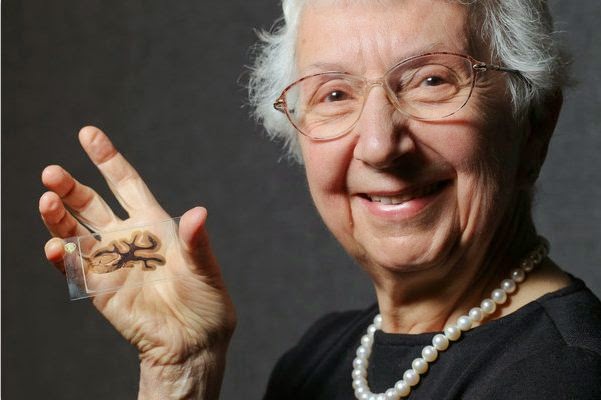
I'm not sure how many in the neuropathology community know this, but our renowned colleague Lucy Rorke-Adams donated slices of Albert Einstein's brain to the Mütter Museum and Historical Medical Library in Philadelphia. Rorke-Adams, a senior neuropathologist at the Children's Hospital of Philadelphia, donated the 46 slices of brain tissue to the museum in 2011. Rorke-Adams had received the slides as a gift from local doctor, who in turn had bequeathed them from his colleague, a neuropathologist who examined the slides on behalf of Thomas Harvey, the man who removed Einstein's brain during an autopsy in 1955. Guess where my first stop will be on my next visit to Philly?

Tuesday, July 8, 2014

Best Post of December 2013: What Happened to Neuropathology in 1946

The next in our 'Best of the Month' series is from December 3, 2013. Since posting, three readers posted comments speculating as to what indeed did happen to neuropathology in 1946. See comments after the post below:

What happened to neuropathology in 1946?

The esteemed Dr. Jim Mandell and his son were recently playing with the amazing Google Books Ngram Viewer. Google Ngram searches a huge corpus of books for the mention of a particular search term. It then graphs the frequency with which that term appears over time. On a whim, the Mandell's entered the search term "neuropathologist". Here's the resulting graph:

I couldn't fit the y-axis label in the picture, but it ranges from 0% up to 0.00000300%. Dr. Mandell challenges his neuropathology colleagues to explain the sharp spike in the usage of "neuropathologist" around 1946-47. Please enter your speculations in the comment section. Dr. Mandell also points out the lamentable fact that it "appears we are past our peak".
______________
Gabrielle Yeaney said...
Not sure if I'm on to something but ...in 1946--certification in pathology was formally recognized, CAP was founded and in 1947--first stereotactic neurosurgical procedure performed. In the early 1940s, AMA recognized pathology as practice of medicine (1943);first neurosurgical training programs and American board of Neurological surgery established. Bernd Scheithauer was born in 1946. Maybe he started publishing at a very early age :) Thanks for the blog
jd said...
JNEN began in 1942.
shipcolldoc said...
It is likely just because WW II was over and a lot of medicine-related terminology other than war trauma would have come to the fore.

Tuesday, December 3, 2013

What happened to neuropathology in 1946?

The esteemed Dr. Jim Mandell and his son were recently playing with the amazing Google Books Ngram Viewer. Google Ngram searches a huge corpus of books for the mention of a particular search term. It then graphs the frequency with which that term appears over time. On a whim, the Mandell's entered the search term "neuropathologist". Here's the resulting graph:

I couldn't fit the y-axis label in the picture, but it ranges from 0% up to 0.00000300%. Dr. Mandell challenges his neuropathology colleagues to explain the sharp spike in the usage of "neuropathologist" around 1946-47. Please enter your speculations in the comment section. Dr. Mandell also points out the lamentable fact that it "appears we are past our peak".

Thursday, June 13, 2013

Best Post of November 2012: Photos reveal unique features of Einstein's cerebral cortex

The next in our "Best of the Month" series is from November 21, 2012:

Photographs taken shortly after his death, but never before analyzed in detail, have now revealed that Einstein’s brain had several unusual features, providing clues about the neural basis of his extraordinary mental abilities.


Nature.com reports that, while doing Einstein's autopsy, the pathologist Thomas Harvey removed the physicist's brain and preserved it in formalin. He then took dozens of black and white photographs of it before it was cut up into 240 blocks. Now, anthropologist Dean Falk of Florida State University in Tallahassee and her colleagues have obtained 12 of Harvey’s original photographs from the National Museum of Health and Medicine in Silver Spring, Maryland, analyzed them, and compared the patterns of convoluted ridges and furrows with those of 85 brains described in other studies. Many of the photographs were taken from unusual angles, and show structures that were not visible in photographs that have been studied previously. The analysis was recently published in the journal Brain. The most striking observation, says Falk, was “the complexity and pattern of convolutions on certain parts of Einstein's cerebral cortex”, especially in the prefrontal cortex, and also parietal lobes and visual cortex.

Tuesday, April 16, 2013

A Rich Focus

The grey arrow is pointing to a Rich focus that has hemorrhaged into the subarachoid space
A Rich focus is a tuberculoma in the cerebral cortex. Rich foci become particularly significant when they rupture into the subarachnoid space and cause tuberculous meningitis. This entity is named for Johns Hopkins pathologist Dr. Arnold Rice Rich (1893-1968), who first described it.

Wednesday, November 21, 2012

Photos reveal unique features of Einstein's cerebral cortex

Photographs taken shortly after his death, but never before analysed in detail, have now revealed that Einstein’s brain had several unusual features, providing clues about the neural basis of his extraordinary mental abilities.


Nature.com reports that, while doing Einstein's autopsy, the pathologist Thomas Harvey removed the physicist's brain and preserved it in formalin. He then took dozens of black and white photographs of it before it was cut up into 240 blocks. Now, anthropologist Dean Falk of Florida State University in Tallahassee and her colleagues have obtained 12 of Harvey’s original photographs from the National Museum of Health and Medicine in Silver Spring, Maryland, analysed them and compared the patterns of convoluted ridges and furrows with those of 85 brains described in other studies.Many of the photographs were taken from unusual angles, and show structures that were not visible in photographs that have been analysed previously. The analysis was recently published today in the journal Brain. The most striking observation, says Falk, was “the complexity and pattern of convolutions on certain parts of Einstein's cerebral cortex”, especially in the prefrontal cortex, and also parietal lobes and visual cortex.

Tuesday, June 19, 2012

Yasser Lopez: A Modern-Day Phineas Gage

Above is an x-ray released yesterday by Jackson Memorial Hospital in Miami, Florida depicting a spear entering the head, traveling through the brain, and piercing the opposite skull surface of 16-year-old Yasser Lopez. The boy is on the road to recovery after being accidentally shot in the head while loading a speargun with a friend earlier this month

"It was about one inch above his right eye straight through," said trauma surgeon Dr. George Garcia. "You could feel the tip under the skin on the posterior part of the skull."

"The most important thing is to resist that temptation to pull the thing out," neurosurgeon Dr. Ross Bullock said. Bullock and his trauma neurosurgeon team determined the tip of the spear inside Lopez's head was actually a screw tip. "It was possible for us to figure out a strategy during the operation to be able to unscrew the tip of the spear, instead of having to get this whole spear dragged out through his brain," Bullock said.

Bullock said Lopez can speak normally. "He can, for example, he says he's not having pain. He's worried about the fact he can't use his left-side properly."

Lopez's story brings to mind that of Phineas P. Gage, whose left frontal lobe was obliterated by a railroad spike that exploded upward when he was tamping it down in 1848.

Phineas P. Gage (1823-1860)
As for the day he got shot, Lopez knows nothing. Doctors expect him to make a near-full recovery.


Read more here: http://www.miamiherald.com/2012/06/18/2856543/doctors-miami-dade-teen-shot.html#storylink=cpy

Neuropathology Blog is Signing Off

Neuropathology Blog has run its course. It's been a fantastic experience authoring this blog over many years. The blog has been a source...