Showing posts with label infectious disease. Show all posts

Thursday, February 4, 2021

Dr. William Yong of UC Irvine co-authors timely update of neuro-COVID

 I recently received an email from the illustrious William Yong, MD at UC Irvine Health. He alerted me to an excellent article which he co-authored entitled Neuropathology of COVID-19 (neuro-COVID): Clinicopathological Update.  Dr. Yong writes: "I don’t normally forward my pubicationss but will do so for this COVID Neuropath review (reviewed over 180 cases from the literature) since things move fast with COVID literature and as Free Neuropathology is a new journal and not in Pubmed yet.  The paper will eventually get into Pubmed as its is NIH funded but process is slower. The article is open access and free to download at Free Neuropathology. If you think it will be of some value to your readers, please feel free to share in your wonderful blog." A PDF version is available here  https://www.uni-muenster.de/Ejournals/index.php/fnp/article/view/2993/3173

Thanks very much, Dr. Yong, for alerting us to this timely update!

Wednesday, March 25, 2020

Lost Smell and Taste Hint COVID-19 Can Target the Nervous System

Matthew Anderson, MD, PhD
Dr. Matt Anderson, chief of neuropathology at Beth Israel in Boston, forwarded me a recent article from The Scientist. The article describes the experience of Alessandro Laurenzi, a biologist working in Bologna, Italy, regarding the proposition that COVID-19 infected patients can have transient loss of the sense of smell. Olfactory sensory loss may play a role in early diagnosis, but it may also help in understanding pathogensis.

Dr. Anderson is quoted in the article, stating that central nervous system involvement by the virus may play a part in the respiratory symptomatology.

Monday, August 12, 2019

Best Post of April 2019: Progressive Multifocal Leukoencephalopathy in patients without obvious immunosuppression

The next in our "Best of the Month" series comes from Friday, April 12, 2019:

I recently received a case in consultation which turned out to be progressive multifocal leukoencephalopathy (PML). Reading the clinical history, it was not entirely clear what predisposed the patient to PML. It wasn't clear, that is, until my mentor (the illustrious BK DeMasters) referred me to a nine-year-old paper by Sarah Gheuens, Gerald Pierone, Patrick Peeters, and Igor J. Koralnik entitled Progressive Multifocal Leukoencephalopathy in individuals with minimal or occult immunosuppression (J Neurol Neurosurg Psychiatry2010;81:247-254). In this series, hepatic cirrhosis -- which was what my patient had -- was among the more common conditions in PML patients with minimal immunosuppression. Other conditions that can be associated with PML in the minimally immunosuppressed are those with renal failure and idiopathic CD4+ lymphopenia,

The authors discuss the possible mechanisms for the development of PML in patients with hepatic cirrhosis:

"[H]epatic cirrhosis can lead to portal hypertension and hypersplenism, with subsequent leucopenia as white blood cells are sequestrated in the enlarged spleen. Furthermore, cirrhosis also leads to hypogammaglobulinemia... It is well known that cirrhotic patients have a higher risk of developing bacterial infections, and 30–50% of deaths among cirrhotic patients are directly caused by infections. Immune dysfunction in hepatic disease may be caused by altered cytokine production, impaired cellular immune response and vascular disturbances, which lead together to increased susceptibility to infections."

Monday, April 15, 2019

PML in a patient with idiopathic CD4 lymphopenia

My last post prompted the inimitable Dr. Murat Gokden to write to me about an interesting case he published in 2015 of a 33-year-old man who with idiopathic CD4 lymphopenia (ICL) who was ultimately diagnosed with progressive multifocal leukoencephalopathy (PML).

Image from Dr. Gikden's paper: A and B are MRI FLAIR images showing hyperintense areas with gyral swelling and sulcal effacement (curved arrows). Image C depicts MR spectroscopy with abnormally elevated choline peak. Image D shows multifocal punctate enhancement in white matter.

Murat Gokden, MD

Friday, April 12, 2019

Progressive Multifocal Leukoencephalopathy in patients without obvious immunosuppression

I recently received a case in consultation which turned out to be progressive multifocal leukoencephalopathy (PML). Reading the clinical history, it was not entirely clear what predisposed the patient to PML. It wasn't clear, that is, until my mentor (the illustrious BK DeMasters) referred me to a nine-year-old paper by Sarah Gheuens, Gerald Pierone, Patrick Peeters, and Igor J. Koralnik entitled Progressive Multifocal Leukoencephalopathy in individuals with minimal or occult immunosuppression (J Neurol Neurosurg Psychiatry 2010;81:247-254). In this series, hepatic cirrhosis -- which was what my patient had -- was among the more common conditions in PML patients with minimal immunosuppression. Other conditions that can be associated with PML in the minimally immunosuppressed are those with renal failure and idiopathic CD4+ lymphopenia,

The authors discuss the possible mechanisms for the development of PML in patients with hepatic cirrhosis:

"[H]epatic cirrhosis can lead to portal hypertension and hypersplenism, with subsequent leucopenia as white blood cells are sequestrated in the enlarged spleen. Furthermore, cirrhosis also leads to hypogammaglobulinemia... It is well known that cirrhotic patients have a higher risk of developing bacterial infections, and 30–50% of deaths among cirrhotic patients are directly caused by infections. Immune dysfunction in hepatic disease may be caused by altered cytokine production, impaired cellular immune response and vascular disturbances, which lead together to increased susceptibility to infections."


Thursday, March 8, 2018

Cerebral malaria in a young woman who had traveled to Africa

A young woman became sick after visiting Zambia. She died a few weeks after onset of headache and flu-like symptoms. The slide is not dirty! The dirty-looking stuff in the picture below is birefringent pigment called hemozoin. This is typical of a falciparum infection. There are ring hemorrhages and Dürck granulomas present as well. Dürck granulomas are accumulations of mononuclear cells, predominantly macrophages, probably related to resorption of ring hemorrhages.


Hemozoin pigment
Dürck granuloma

Saturday, September 17, 2016

CAP16 Abstract Highlights - Naegleria fowleri: Understanding the Clinical Presentation and Autopsy Findings of a Rare and Almost Universally Fatal Central Nervous System Infection

The 2016 annual meeting of the College of American Pathlologists (CAP16) is coming up September 25-28 in Las Vegas. In this series of posts, I'll be featuring poster abstracts of particular interest to neuropathologists.

Alexander T. Damron and colleagues at Baylor College of Medicine in Houston discuss Naegleria fowleri CNS infection in Poster #114:


Naegleria fowleri is a free-living ameba known to cause primary amebic meningoencephalitis (PAM). Moreover, PAM is an acute, fulminating, and hemorrhagic infection that occurs in healthy young children with fresh water exposure in warm climates. It is postulated that Naegleria fowleri enters through the nasal passages and crosses the cribriform plate, where it reaches the subarachnoid space and disseminates into the olfactory lobes. Visvesvara et al (2007) performed a retrospective study of all reported N fowleri infections in the United States from 1937 to 2013 and found 3 survivors in 142 reported cases. Only 27% of the 142 cases were diagnosed before patient death. We present a case of a previously healthy 14-year-old boy who presented with fever, headache, vomiting, and altered mental status 8 days after swimming in a warm freshwater lake. Cerebrospinal fluid studies showed organisms consistent with amoeba (Figure 265, C). Despite neuroprotective measures and antimicrobial medications, the patient was pronounced brain dead 9 days after admission. Autopsy revealed the cause of death to be PAM from infection with N fowleri with cerebral edema and tonsillar herniation (Figure 265, D). Microscopic examination of the central nervous system revealed amoebic organisms infiltrating the meninges and diffusely involving the brain parenchyma in a perivascular distribution (Figure 265, A and B). The most critical aspect in treating patients with PAM is early detection and prompt initiation of multiple antimicrobials and neuroprotective measures.Despite current recommendations, the high mortality rate of these infections (97%–98%) suggests that an effective treatment for PAM is not yet known.

Thursday, August 11, 2016

My all-time favorite post ever: Whither the Illusory Cowdry B Inclusion?

I have put up a grand total of 619 blog posts since this site got started back on October 31, 2007. I was recently asked which was my favorite. No question, my all-time favorite is about the illusory Cowdry B body. This post has even made it into the references for the Wikipedia page on Cowdry bodies. By the way, the reward of a 10 dollar bill which I posted six years ago to the person who sends me a picture of this structure still stands! I have no fear of being parted from my Jefferson as, in my opinion, the Cowdry B body is the Sasquatch of neuropathology. Here's the post from November 13, 2008:

In a recent post about poliomyelitis, the illustrious Dr. John Donahue of Brown University (pictured) correctly pointed out that I did not mention the presence of Cowdry B inclusions in my histological description of the disease. Having never seen polio under the microscope, I went looking in textbooks and on the web for a photomicrograph of a polio-related Cowdry B inclusion. Failing in my search, I turned to the esteemed Dr. Tom Smith of the University of Massachusetts to see if he had such a picture. With his permission, I quote Dr. Smith’s email to me:

“I have the same problem you and everyone else seems to have -- I've never seen one myself and cannot find any photos of one either in my own file or anywhere in books or on the web. I remember reading a description a long time ago - I think it might have been from the old AFIP neuropath teaching slide set - that they were supposed to be small eosinophilic (?) nuclear inclusions that were sometimes seen in neurons in poliomyelitis. I don't remember actually seeing them in the slide in that teaching set. From the description, I had the impression they might have resembled Marinesco bodies or maybe even normal but prominent nucleoli... or perhaps those small inclusions seen in some neurodegenerative dementias. They don't seem to be as 'specific' as Cowdry A inclusions and perhaps they don't even exist? Frankly I think Cowdry B inclusions have reached the point where they should be relegated to the trashbin of neuropathology.” (Emphasis added.)

I recently photographed a Marinesco body (see picture, arrow points to the eosinophilic body) within the 
nucleus (outlined) of a pigmented neuron in the substantia nigra. Could the Cowdry B inclusion be an elaborate hoax perpetrated upon us by Dr. E.V. Cowdry when he first described Cowdry type A and type B inclusions in 1934? Dr. Smith’s response:
“Well, I doubt that it was a hoax but I think some of those folks were quite prone to seeing things that just the passage of time (and new information) has proved to be ‘not real’. Another case in point - how many Alzheimer type 1 astrocytes have you
seen?”

I then wrote back to the individual who got me started on this hunt for the illusory Cowdry B in the first place: John E Donahue, MD. Here’s what Dr. Donahue had to say about the issue:
“I think these descriptions are very old and go back to the day where everything was described visually, without knowing etiology. (Remember, there are eight structures of Scherer from the original 1938 article, only three of which are really relevant anymore, and maybe even the gliosarcoma being a tertiary structure may be going the way of the dinosaur since I've heard recently that the gliosarcoma arises from a single precursor cell)…”

Fuller and Goodman, in Practical Review of Neuropathology (Lippincott, 2001)define a Cowdry B purely on morphological grounds -- with no implication as to the cause (polio or otherwise) -- as being small, eosinophilic, intranuclear inclusions with no halo and causing no nuclear effacement. I quote page 20: “[T]he quotidian Marinesco bodies that are routinely observed in the neurons of the pigmented brainstem nuclei are sterling examples of the Cowdry type B beau ideal.”

Perhaps the Marinesco body, rather than a “sterling example”, may in fact be the only example of a Cowdry B inclusion! Come to think of it, I think I’ll offer a $10 reward for anyone who can send me a photomicrograph of a polio-related Cowdry B inclusion. Email me the photo at brian.moore@ucdenver.edu

I hope you have better luck finding one than I did!

Monday, July 18, 2016

A 60-year-old man presents with acute confusion, aggressive behavior, and aphasia


T2-weighted FLAIR MRI 

A 60-year-old man presents with acute confusion, aggressive behavior, and aphasia. Imaging reveals a heterogeneously enhancing mass (5 x 2 x 2 cm) in the left medial temporal lobe with extensive peripheral edema (image above), but no significant contrast enhancement. A subtotal temporal lobectomy was performed on suspicion of a low-grade glioma.


High-power view of biopsy from left temporal lobe

The diagnosis is herpes simplex encephalitis. PCR amplification of DNA from the specimen more specifically identified the causative agent as herpes simplex virus, type 1 (HSV1). The most common identifiable cause of viral encephalitis in hospitalized patients, HSV1 classically exhibits tropism for the orbitofrontal and temporal lobes bilaterally, but can on occasion present as a unifocal mass mimicking neoplasm as exemplified in this case. Untreated cases have a mortality rate of up to 70%, while in patients treated with a 14- to 21-day course of acyclovir the mortality rate is less than 10%. Although the patient in this case clinically improved while on a four-week course of acyclovir, he did not return to baseline functioning and was discharged to a nursing home.

Primary infection with HSV1 is usually subclinical. Symptomatic primary infection may manifest as gingivostomatitis, pharyngitis, or a mononucleosis-like syndrome. During the initial infection, the virus undergoes retrograde transport along sensory axons and becomes established in latent form in sensory ganglia, especially the trigeminal ganglion. Reactivation of latent virus produces recurrent mucocutaneous lesions most commonly involving the vermilion border of the lips (herpes labialis) that are recognized as cold sores or fever blisters. About one-third of herpes encephalitis cases are associated with primary infection, while the remainder result from reactivation of latent virus. It remains unclear whether viral reactivation occurs within the brain itself (herpes simplex viral DNA has been detected in normal brain) or in peripheral sensory ganglia, such as the trigeminal ganglion, with secondary axonal transport to the CNS. Olfactory pathways are also likely routes of retrograde viral transport. In contrast, HSV2 infection is more commonly encountered in newborns, acquired via genital infection from the mother during vaginal birth.

A hypercellular specimen with reactive astrocytes and occasional mitotic figures can raise the specter of astrocytoma. However, demonstration of extravasated blood and ischemic necrosis with infiltration by neutrophils, lymphocytes, and CD68-positive macrophages leads to a diagnosis of hemorrhagic necrotizing encephalitis.  Although the presence of haloed Cowdry A viral inclusions provide diagnostic reassurance (see photomicrograph), they are not always seen and are not specific to HSV. Immunohistochemistry for HSV1/2 is positive in infected cells.

Diagnosis:  Herpes simplex virus type 1 (HSV1) encephalitis

Monday, June 27, 2016

Best Post of April 2016: Zika virus isolated from fetal brain tissue

The next in our "Best of the Month" series is a post from April 8, 2016:

Cheng-Ying Ho, MD, PhD
Dr. Cheng-Ying Ho, neuropathologist at Children's National Medical Center in Washington DC, recently authored a report in the New England Journal of Medicine entitled Zika Virus Infection with Prolonged Maternal Viremia and Fetal Brain Abnormalities. In this report, Dr. Ho and colleagues describe a case of a pregnant woman and her fetus infected with the Zika virus during the eleventh week of gestation. The fetus had a significantly decreased head circumference by the twentieth week of gestation. Given the grave prognosis, the mother elected to terminate the pregnancy at 21-weeks gestation. Postmortem analysis of the fetal brain revealed diffuse cerebral cortical thinning, a high Zika virus RNA load, detection of viral particles, and isolation of the Zika virus from brain tissue. This is the first report of Zika virus isolation from fetal brain tissue. This finding fulfills Koch's second postulate regarding the isolation of a pathogen from a diseased organism and therefore goes a long way toward strengthening the association between congential Zika virus infection and fetal brain damage. This is an important work from a rising star in the neuropathological firmament. As a fellow neuropathologist commented, Dr. Ho is "a very talented young investigator from whom you will be hearing a lot in the future".

Tuesday, April 19, 2016

Photomicrographs of Zika-infected fetal brain

Dr. Cheng-Ying Ho, neuropathologist at Children's National Medical Center in Washington DC kindly provided the photomicrographs below (click to enlarge), which were published with her important case report about which I recently published a blog post.
Thinning (left) with apoptosis (inset left) as compared to relatively spared cerebral cortex (right)
The legend for these pictures is as follows: "
In postmortem analyses of samples obtained from the fetus, an area of parietal cortex has abundant apoptotic neurons (Panel A), with detail shown in the inset view. The unaffected occipital cortex is thicker than the parietal cortex (Panel B), as indicated by the vertical bars." 

Tuesday, April 12, 2016

"Funny worm-like profiles" in a meningioma

Howard Chang, MD, PhD
The illustrious Dr. Howard Chang of Michigan State University recently sent me photomicrographs of a meningioma specimen from the posterior fossa of a 63-year-old female. Within the meningioma were noticed "funny worm-like profiles". The specimen was sent to the Centers for Disease Control, which provided reassurance that these profiles were not parasites. The inimitable Dr. John Donahue wondered whether this was simply embedded ependymal lining. I wondered the same thing. Dr. Chang tells me that GFAP was not ordered by the general pathologist signing out this case. Meanwhile, the eminent Dr. Mark Cohen felt -- despite the CDC's pronouncement -- that the structures did not look human. Please help our colleague Dr. Chang by making any diagnostic suggestions in the comments section.


Friday, April 8, 2016

Zika virus isolated from fetal brain tissue

Cheng-Ying Ho, MD, PhD
Dr. Cheng-Ying Ho, neuropathologist at Children's National Medical Center in Washington DC, recently authored a report in the New England Journal of Medicine entitled Zika Virus Infection with Prolonged Maternal Viremia and Fetal Brain Abnormalities. In this report, Dr. Ho and colleagues describe a case of a pregnant woman and her fetus infected with the Zika virus during the eleventh week of gestation. The fetus had a significantly decreased head circumference by the twentieth week of gestation. Given the grave prognosis, the mother elected to terminate the pregnancy at 21-weeks gestation. Postmortem analysis of the fetal brain revealed diffuse cerebral cortical thinning, a high Zika virus RNA load, detection of viral particles, and isolation of the Zika virus from brain tissue. This is the first report of Zika virus isolation from fetal brain tissue. This finding fulfills Koch's second postulate regarding the isolation of a pathogen from a diseased organism and therefore goes a long way toward strengthening the association between congential Zika virus infection and fetal brain damage. This is an important work from a rising star in the neuropathological firmament. As a fellow neuropathologist commented, Dr. Ho is "a very talented young investigator from whom you will be hearing a lot in the future".

Monday, March 21, 2016

Cornea of an elderly female soft contact lens wearer afflicted by acanthamoeba keratitis

Evacuated cyst walls embedded in corneal stroma
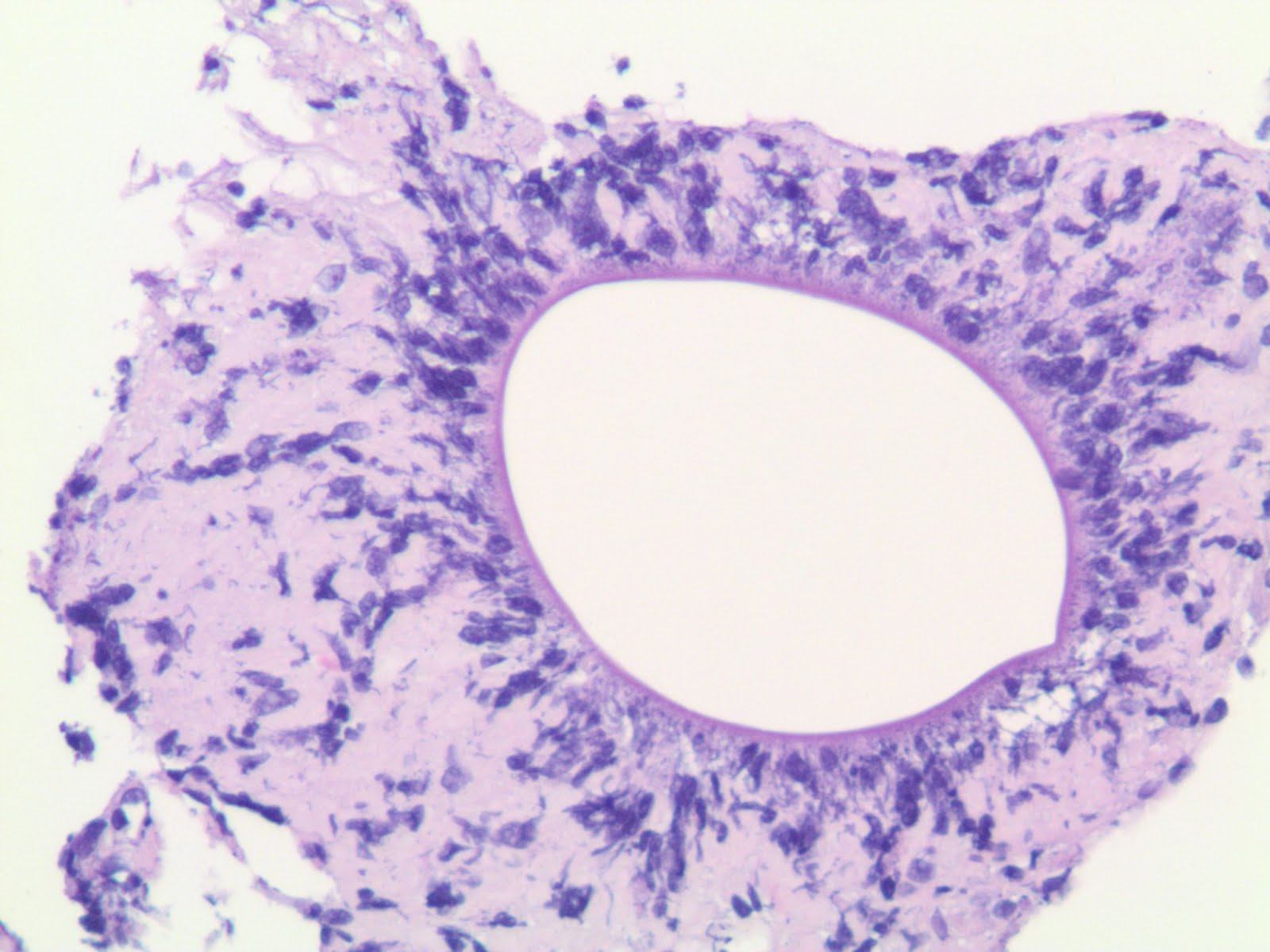
Acathamoeba keratitis classically afflicts soft contact lens wearers who use contaminated homemade saline solutions or wear their contact lenses while swimming or in hot tubs. Corneal infection is usually very painful. In some medically intransigent cases, as exemplified in this case, corneal transplantation may be necessary. The chitinous wall of the organism is oval, while the cytoplasm typically retracts from the cyst wall in tissue sections. The cyst contains a round nucleus with a distinct nucleolus.
Acanthamoeba nucleus with distinct nucleolus

Wednesday, September 25, 2013

Thirteen patients in New England put at risk for iatrogenic CJD

CNN reports that a patient who had undergone neurosurgery in New Hampshire later developed autopsy-confirmed sporadic Creutzfeldt-Jakob disease (sCJD). Before the patient's disease was discovered, the same nonsurgical equipment used on the CJD patient was used on thirteen subsequent patients, putting those patients at risk for prion infection. The Centers for Disease Control has said that no cases of the disease linked to the use of contaminated medical equipment have been reported in the United States since 1976.

Wednesday, September 11, 2013

Best Post of April 2013: A Rich Focus

The next in our "Best of the Month" series is a post from April 16, 2013.

The grey arrow is pointing to a Rich focus that has hemorrhaged into the subarachoid space
A Rich focus is a tuberculoma in the cerebral cortex. Rich foci become particularly significant when they rupture into the subarachnoid space and cause tuberculous meningitis. This entity is named for Johns Hopkins pathologist Dr. Arnold Rice Rich (1893-1968), who first described it.

Tuesday, April 16, 2013

A Rich Focus

The grey arrow is pointing to a Rich focus that has hemorrhaged into the subarachoid space
A Rich focus is a tuberculoma in the cerebral cortex. Rich foci become particularly significant when they rupture into the subarachnoid space and cause tuberculous meningitis. This entity is named for Johns Hopkins pathologist Dr. Arnold Rice Rich (1893-1968), who first described it.

Monday, October 1, 2012

A 32-year-old HIV+ male presenting with mental status changes

 Soon after admission, the patient lapsed into a coma and died.

Minimal edema (brain weight: 1500 grams)

Some hypoxic-ischemic damage as evidenced by Purkinje red neuron change
"Soap bubble" changes around some vessels (sample from basal ganglia)
Giant cells among mixed inflammatory infiltrate in the meninges
Inflammation appears to extend minimally into brain parenchyma
Organisms consistent with crytopcoccus neoformans amid inflammation
Mucicarmine-positive fungi capsules highlighted in red, with some capsules partially deficient
Diagnosis: Cryptococcal meningoencephalitis with early hypoxic-ischemic encephalopathy.

Neuropathology Blog is Signing Off

Neuropathology Blog has run its course. It's been a fantastic experience authoring this blog over many years. The blog has been a source...