Friday, December 16, 2011

Naegleria-tainted neti pots possibly killed two people

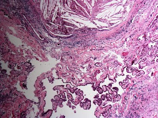
Naegleria fowleria (photo courtesy of Dr. Mark Cohen)
Now that it's winter and our noses and sinuses are being dried out by our heating systems, many of us turn to neti pots (also known as 'nose bidets'). The illustrious J. Clay Goodman, MD, neuropathologist at Baylor, just sent me an article from The Houston Chronicle warning people to use distilled, bottled, or boiled water. Officials in Louisiana are investigating whether a 51-year-old woman and a 20-year-old man both contracted the brain-eating amoeba Naegleria fowleri after using tap water in their neti pots. The disease, while rare, is most commonly contracted after inhaling water from a lake, pond or river. If investigators confirm the two victims died after using tap water from a neti pot it will be the first time the disease has been contracted from tap water.

Wednesday, December 14, 2011

Brigham and Women's Hospital Names Neuropathologist as New Chairman

Dr. Jeffrey A. Golden
In an obvious attempt to keep up with Massachusetts General Hospital, whose pathology chief is neuropathologist Dr. David Louis, Brigham and Women's Hospital in Boston has just announced that another neuropathologist, Dr. Jeffrey Golden, will become chair of pathology at that institution. Effective March 1, 2012, Dr. Golden will become the Ramzi S. Cotran Professor of Pathology at Harvard Medical School.  Brigham President Betsy Nabel, MD had this to say in a statement released today: Dr. Golden comes to BWH "from The Children's Hospital of Philadelphia, where since 2008, he has been serving as Pathologist-in-Chief in CHOP's Department of Pathology and Laboratory Medicine, cited as one of the leading pathology and developmental biology programs in the U.S. and one of the top NIH-funded research programs in Pathology.... Dr. Golden is recognized as an outstanding clinician, a highly skilled educator focused on training the next generation of physician/scientists in pathology and developmental biology, and a cutting-edge basic and translational research leader.  His studies in the multiple aspects of developmental biology and early nervous system development are key to gaining a greater insight into the pathogenesis of human diseases, including brain malformations, mental retardation, epilepsy and autism.  His efforts to date of establishing and elevating the bench to bedside and back to bench paradigm in the fast changing field of developmental biology and molecular diagnostics reflects a true commitment to quality and excellence in the application of new knowledge at the front lines of patient care."

As one neuropathologist commented to another: "Apparently neuropath is now ruling the world."

Monday, December 12, 2011

Improvements Recently Made to the AANP Website

The website for the American Association of Neuropathologists has recently undergone some improvements that might be of interest to NeuropathologyBlog readers:

The membership directory should be changed so it is searchable by location (hospital, city, or state), as well as by name.

- The Education Section provides useful links for those of us (myself included) who need to get our SAM credits in to the American Board of Pathology for maintenance of certification.

- A new tab has been added under the Professional Affairs heading, entitled “Survey". Results of recent AANP surveys are posted here.

A new heading for the left hand column on the home page has been added entitled “Links of interest". Several links pertinent to neuropathologists are listed. (Hey, where's NeuropathologyBlog?!?)


Thanks to AANP Vice-President Elect Liz Cochran, MD for providing me with this information.

Sunday, December 4, 2011

Best Post of June 2011: A 58-year-old woman with headache and unsteadiness of gait

The next in our "Best of the Month" series is from June 14, 2011:

Here's a recent case of mine involving a lesion in the third ventricle of a 58-year-old woman who presented with headache and unsteadiness of gait. First, the imaging:


Axial CT
Sagittal T1 MRI

Axial T2 MRI
Coronal Gradient Echo
The neuroradiologist thought perhaps the lesion was a craniopharyngioma. But, the neurosurgeon was surprised that the lesion seemed to "pop out". Here's the specimen I received:
Outer Surface

Cut Surface
I smeared some of the viscous fluid from the cut surface onto a glass slide, cover-slipped it, and viewed it under polarized light:


Those cholesterol crystals certainly seem to suggest the possibility of craniopharyngioma. But, craniopharyngiomas do not have a smooth outer surface and do not just "pop out" into the neurosurgeon's hand. Here was my first glimpse of the histology the next day:
Cholesterol clefts and giant cells. Note in the upper picture that this xanthogranulomatous reaction appears to be continuous with the choroid plexus. Could this be a xanthogranuloma of the choroid plexus? I considered that, until I looked elsewhere on the slide and saw this:


This is the ciliated epithelium characteristic of a colloid cyst of the third ventricle, a tumor that can indeed "pop out" into the neurosurgeon's hand. Gross inspection did not reveal a cyst per se because the exuberant xanthogranulomatous reaction obliterated it. Cases like this have been described in the literature. For example, Dr. David Louis and colleagues wrote in a 1994 article entitled Third ventricle xanthogranulomas clinically and radiologically mimicking colloid cysts: Report of two cases (Journal of Neurosurgery. 81(4):605-9, 1994 Oct.) that "histopathologic examination revealed xanthogranulomas of the choroid plexus with only microscopic foci of colloid cyst-like structures." I would argue quite the opposite: that the xanthogranuloma derived from the colloid cyst and extended into the choroid plexus. Indeed, Dr. Peter Burger and colleagues write in the fourth edition of Surgical Pathology of the Nervous System and Its Coverings that "a xanthogranulomatous reaction occasionally supervenes in colloid cysts, largely replacing the epithelium in some cases."

Cool case.

Friday, November 18, 2011

Neuropathology Focus of New SNO President's Address Today


Over 1400 attendees at 2011 SNO meeting in Anaheim

                   Dr. Aldape discussed brain tumor biomarkers                
Same view as above, at 40X
Neuropathlogy took center stage at the Society for Neuro-Oncology (SNO) meeting today as Dr. Kenneth D. Aldape, neuropathogist at the University of Texas MD Anderson Cancer, was introduced as the next president of the society. Dr. Aldape also delivered an address to more than 1400 attendees about the future of surgical neuropathological reporting. Aldape's research centers on the clinical utility of brain tumor biomarkers as prognostic indicators. He described how the use of biomarkers will change the way surgical neuropathology diagnoses are rendered. He noted that the broad morphologic spectrum one sees in gliomas makes the recommendations of the World Health Organization sometimes difficult to implement. For example, the difference between a WHO grade II and grade III astrocytoma is based principally on whether or not mitotic figures are "brisk" as interpreted by the examining neuropathologist. Yet, the imprecision of that approach is obvious when one considers variables such as the diligence of the neuropathologist in identifying mitotic figures, the often equivocal morphology of apoptotic-versus-mitotic cells, as well as the variable technical quality of tissue processing and staining. The use of biomarkers will make diagnoses more reliable and will provide more useful prognostic information to the oncologist. Dr. Aldape projected onto the screen an example of a recent surgical pathology report he had generated at MD Anderson. The report highlighted the status of the several biomarkers, including G-CIMP, IDH1, pHH3, MIB1, 1p/19q deletion, and MGMT. Then, only in the last sentence of the comment, was it noted that the histology and biomarker profile was most consistent with a WHO grade III (anaplastic) astrocytoma. It was as if the WHO diagnosis and grade were a perfunctory afterthought. Finally, Aldape noted that biomarkers will become even more important in the coming years as biomarker data becomes "actionable", allowing the oncologist to personalize the treatment of a particular tumor depending on the biomarker profile. These are exciting times....

Thursday, November 17, 2011

Kois, Perry, Giannini, and Zagzag Among Neuropathologists at SNO Meeting

Neuropathologists are well represented at the Society for Neuro-Oncology (SNO) meeting going on here in Anaheim, California. Today I ran into Dr. Arie Perry of UCSF and was pleased to make the aquaintence of Dr. Nancy Kois of St. Alphonsus Hospital in Boise, ID.


I had a chance to catch up with the Mayo Clinic's Dr. Katerina Giannini who is in attendance this weekend. I also just ran into NYU's Dr. David Zagzag, who confirmed that his institution is actively seeking a neuropathologist. Anyone interested in the position can email him at dz4@nyu.edu.

Wednesday, November 16, 2011

SNO is coming down in Southern California!!

For the next couple of days, I'll be blogging live from the 16th annual meeting of the Society for Neuro-Oncology (SNO) in Anaheim, California. SNO is the most inter-disciplinary of the neurologic societies, with membership that includes neurosurgeons, neuro-oncologists, neuropathologists, radiation oncologists, neuroradiologists, pediatricians, laboratory scientists, nurses, and other specialists. According to a report from SNO's membership committee, a full 5% of SNO membership comes from the specialty of pathology. I am among that 5%. SNO's acknowledgement of the pivotal role neuropathology plays in brain tumor research and treatment is exemplified by the fact that this year the society has recognized a neuropathologist -- Kenneth Aldape, MD from the University of Texas MD Anderson Cancer Center -- as recipient of the Victor Levin Award in Neuro-Oncology Research. I'll be in attendance on Friday when Dr. Aldape receives the Levin Award and delivers a plenary session on tumor genetics. SNO has further recognized neuropathology by including "pathology" among the nine Young Investigator Round Table career interest luncheons. Finally, in reviewing the abstracts from the meeting, I see that several posters include neuropathologists among the authors. Although most of these authors are placed in other specialty categories in the abstract book, there is actually a distinct section devoted specifically to pathology. The senior author of one of the posters in the pathology section is a neuropathologist whom I have described previously in this blog as a "rising star in the neuropathology firmament": Craig Horbinski, MD, PhD.  Dr. Horbinski's abstract is  entitled Glioblastoma Survival Varies According to Degree of EGFR Amplification.

The outgoing president of SNO, Frederick F. Lang, MD, writes in the latest issue of SNO News that one of the goals of his presidency has been to "extend a cooperative hand to other like-minded organizations in the USA and internationally. The wisdom of SNO is its inclusion of multiple disciplines, and I have always thought that other organizations should benefit from this wisdom."  In the spirit of this multidisciplinary approach, SNO has partnered this year with the Section of Tumors of the American Association of Neurological Surgeons (AANS) and Congress of Neurological Surgeons (CNS). I would urge the American Association of Neuropathologists (AANP) to consider partnering with SNO to create a joint meeting. A productive cross-pollination would undoubtedly result.

Keep reading this blog over the next couple of days as I report on the interesting educational and research presentations that will occur as SNO comes sunny Southern California!

Thursday, November 10, 2011

MD/PhD Student Seeks Advice About a Career in Neuropathology


I recently received an inquiry from a medical student named Stephen Briggs. Rather than pontificate on this topic myself, I am posting, with his permission, Stephen's email with hope that the readership of this blog might offer some advice to this young man in the comment section of this post. Thanks in advance to anyone who would like to put in their two cents' worth.

Stephen Briggs
Dear Dr. Moore,
  I have been avidly following your blog for a while now, and I would like to ask you a few questions about the field of neuropathology. I am an MD/PhD student, currently in the PhD phase of my training. I am researching neurological disease, and I want to continue in this field. I am still relatively early in the program (I just finished the first year of my PhD), but I would like to start thinking about possible future directions. Neuropathology seems to be a great fit based on my interests, but I would like to discuss some of the details with you.
  First, you have mentioned in some posts that a neurology residency followed by neuropathology is no longer the standard pathway (September 2010 guest post). Is it actually a disadvantage? If I want to do neuropathology work, should I plan to go through pathology? The guest poster (Dr. John Donahue) seemed to think that the neurology to neuropathology was a long shot for new students, because most departments want a more general pathologist instead of a total specialist in neuropathology.
  Second, I was interested in your take on research in neuropathology. To an outsider, it seems particularly well suited: it includes predictable work hours, access to human tissue, and a generally academic mindset. Is the actual practice different? Are there any challenges to pathology research that I may not be aware of? If I chose to do research in neurology instead, would it be substantially different?
  Finally, I am interested in the long-term prospects of the field. I count fully 20 openings posted on your blog, which certainly seems hopeful. However, from searching on Google I have seen horror stories about how difficult it can be to get into the field of pathology in general. Some pathologists report taking fellowship after fellowship to avoid being unemployed while waiting for a position to open up. Also, some residents were worried about the field as a whole, as it is easier to computerize or outsource than other fields of medicine. Are these concerns justified?
  Thank you for maintaining your blog. I am glad that there is a resource for students to find out a little about neuropathology, and to feel connected to the field as it evolves.
  Stephen Briggs

Sunday, November 6, 2011

Best Post of May 2011: The Timeless Dr. Adelman

The next in our "Best of the Month" series comes from May 20, 2011:

Lester S. Adelman, MD
I was recently sent this 1992 essay written by Dr. Lester S. Adelman, retired neuropathologist at Tufts New England Medical Center. It amusingly illustrates the anonymity in which we pathologists toil:

Pathologists spend their time in the figurative,
and sometimes literal bowels of the hospital.
Even those of us who specialize in the pathology
of the brain occupy this nether region. The
patients who benefit from our brilliant diagnoses
are often as little aware of us as they are of
the hospital laundry.
This life of anonymity has its rewards. Frozen sections
are only rarely done on nights and weekends, and straightforward
cases are disposed of quickly. The intellectual and
visual pleasures of the job are great. Every once in a while,
however, we are reminded of the fact that we have given up
a part of doctoring, the part that has to do with knowing the
gratitude of the patients whom we help.
Some years ago, in the era before CT scans and MRIs,
a patient was admitted to our hospital with a brain tumor.
Preoperative radiologic diagnoses in those days were based
on angiography, and this patient's angiogram strongly suggested
a glioblastoma multiforme. The patient was given the
bad news, and a biopsy was done to confirm the diagnosis.
The surgeons also thought the tumor was a glioblastoma,
but when I looked at the frozen section, I discovered the
tumor was clearly a meningioma. With this diagnosis, the
surgeons were able to find a plane of dissection around the
tumor and remove it completely.
I was happy with the outcome and thought I had accepted
the fact that the patient would never know my part .
in it. It was not long after that I was to come face-to-face
with the shortcomings of my chosen medical specialty.
A week later I was at a conference with the neurosurgeons
when I noticed that one of the residents was wearing
an expensive new wristwatch. A moment later I noticed that
a senior neurosurgeon had a similar watch on his wrist. A
quick check of the audience revealed that they all had new
watches!
"What's going on?" I asked.
"You remember Mr. X, the patient with the meningioma?
He's a jeweler, and he was so happy he didn't have
a glioblastoma that he gave us all new watches."
"How about me?" I whined. "I'm the one who cured
him. If it weren't for me, you would have quit, thinking he
had a glioblastoma."
The physicians smiled graciously and conceded this was
probably the case.
Years have passed, and despite the advances in imaging,
my neurosurgical colleagues continue to consult me about
diagnoses. But when I want to know what time it is I still
consult my Timex.

Friday, October 28, 2011

Dr. Doug Anthony becomes Chief-of-Pathology at Brown-affiliated hospitals

Excerpts from a message from Timothy J. Babineau, MD, President and Chief Executive Officer, Rhode Island Hospital and The Miriam Hospital

We are pleased to announce the appointment of Douglas C. Anthony, MD, PhD, as chief of pathology at Rhode Island Hospital and The Miriam Hospital, effective February 1, 2012. A neuropathologist, Dr. Anthony will lead the clinical, educational and research pathology programs for Lifespan, as well as help to bridge pathology and the neurosciences through the Norman Prince Neuroscience Institute.


Douglas C. Anthony, MD, PhD
Dr. Anthony comes to Rhode Island Hospital from the University of Missouri, where he is the chair of the department of pathology and anatomical sciences and professor of neurology.  At the University of Missouri Health Care he serves as chief of pathology and medical director of pathology clinical laboratories. He also serves as a pathologist at the Harry S. Truman Veterans Administration Medical Center, Women and Children’s Hospital, and the Ellis Fischel Cancer Center, and as a neuropathologist for the Office of the Medical Examiner in Boone, Callaway and Greene counties in Missouri.  Previously, he was on the faculty at Harvard Medical School and served as a neuropathologist at Boston Children’s Hospital, Brigham and Women’s Hospital, and as a neuropathologist consultant to the Office of the Chief Medical Examiner for the Commonwealth of Massachusetts.

He has won numerous awards, including the Dr. Edison H. and Sallie Y. Miyawaki Teaching Award in Neurosciences at Harvard Medical School; the Excellence in Education Award in pre-clinical sciences at the University of Missouri; and the Order of Socrates award at the School of Medicine at the University of Missouri.

Dr. Anthony’s appointment further demonstrates our commitment to becoming a leader in the neurosciences, and we are confident that he will be a tremendous asset to our clinical team.  Please join me in welcoming and congratulating Dr. Anthony.

Wednesday, October 26, 2011

NIH Loan Repayment Program a godsend for young neuropathologists interested in research

Today I feature a guest post from the illustrious Dr. Mike Lawlor.

Hi Everyone,

I'd just like to tell everyone about a program that the NIH offers, which may be of great interest to Neuropathology Fellows, Research Fellows, and Junior Faculty.  It's called the NIH Loan Repayment Program (LRP), and it's designed to encourage people with an interest in research to remain in the academic research environment.  From what I've heard, the NIH created this program so that people with lots of student loans (mostly medical doctors) with an interest in research would not give up their research careers due to their loan debt.
I just got an email from the LRP people, and they now have a webinar available to help people apply.  The link for the webinar is: http://bit.ly/nihlrptutorial 

Michael W. Lawlor, MD, PhD
The award offers up to $35,000 per year for up to two years, and you're able to apply for renewals whenever it runs out.  This counts as income, but the NIH also pays the federal income tax on this grant for you.  I ended up owing about $2000 in state tax every year for having the award, but that doesn't seem like much as you watch your student loans disappear.

Here's what you'll need:

1) The grant application, which can be found this site.  The forms aren't too labor-intensive, and you need to propose a 2-year research program and be able to guarantee a 50% effort commitment to the research.

2) A bunch of info on your student loans, most/all of which can be found on your monthly statements.  Your student loan burden needs to exceed a certain amount before you qualify, and the amount you need is dependent on your income.  Given the non-stellar income of most neuropath fellows and research fellows, it's actually pretty easy to qualify on financial grounds.  You may continue to apply for renewals as long as you continue to meet the eligibility requirements.

3) Someone to vouch for you.  Your research adviser will need to reply to an email that confirms that you are devoting 50% effort to your research every quarter.  After that confirmation is made, and you confirm that the prior payment made it to the right account, they apply a payment to your educational loans.

Anyway, I've been in this program for the past 2 years, had it renewed for another year, and am in the process of writing another renewal.  It really is a fantastic program with excellent support.  The next deadline is November 15th, so there's definitely time for you guys to put something together.  Once you submit the application, you'll hear nothing for about 6 months, and then they'll ask you for some updated student loan information if you've made it through their scientific review process.  The application cycle is once per year.

I hope that this helps some of you, and good luck!

Thanks for this helpful information, Mike!

Monday, October 17, 2011

Treatable Neurological Disorders Misdiagnosed as Creutzfeldt-Jakob Disease

A case of primary CNS angiitis thought to be sCJD
Dr. Mark Cohen and a team of workers at Case Medical Center in Cleveland, Ohio have published an important article in the Annals of Neurology entitled Treatable Neurological Disorders Misdiagnosed as Creutzfeldt-Jakob Disease (Ann Neurol 2011;70:437–444). Why is this article important? Well, because mistaking a survivable, treatable disorder for a fatal, non-treatable disorder is not optimal. Cohen's team reviewed the pathologic diagnoses of 1,106 patients who were referred for potential prion disease to the National Prion Disease Pathology Surveillance Center (NPDPSC) at Case Western Reserve University from 2006 to 2009. About one-third of the cases did not have prion disease, with Alzheimer disease and vascular disease being the most common conditions accounting for dementia. Further, about one-quarter of the non-prion cases had treatable diseases, including immune-mediated disorders, neoplastic disorders such as lymphoma, as well as infectious and metabolic disorders. The immune-mediated disorders included primary angiitis of the CNS, acute disseminated encephalomyelitis, limbic encephalitis, neurosarcoidosis, paraneoplastic cerebellar degeneration, and one case of Wegener granulomatosis. Of note, more than half of patients with a treatable dementia had a positive CSF 14-3-3 protein test, highlighting the danger of relying too heavily on this test in making a diagnosis of sporadic Creutzfeldt-Jakob disease (sCJD). It turns out that the most specific test for distinguishing CJD from other diseases was magnetic resonance imaging. Drs. David Perry and Michael Geschwind, in a review of this study in the September 2011 issue of Nature Reviews Neurology, (Perry, D. C. & Geschwind, M. D. Nat. Rev. Neurol. 2011:7, 479–480) write: "The fact that many of the non-prion diagnoses in the present study were potentially treatable RPDs [rapidly progressive dementias] should prompt thorough diagnostic testing in patients who are suspected of having sCJD, in order to rule out mimics. The use of CSF testing, contrast-enhanced MRI, and autoimmune antibody screening is supported by this study."

Monday, October 10, 2011

Attractive Neuropathology Postdoc Position in Neurodegenerative Disease Now Available at UCSF

Lea T. Grinberg, MD, PhD.
I recently received an email from Lea T. Grinberg, MD, PhD, a neuropathologist originally from Sao Paulo, Brazil now working at UCSF. She is recruiting a postdoc to work with her. Dr. Grinberg specializes in aging and neurodegenerative diseases and was recently awarded an R01 grant to study early stages of AD and FTLD-TDP in postmortem human tissue. She has access to a large brain bank associated with the Brazilian Aging Brain Study Group as well as the UCSF Memory and Aging Center brain bank. The postdoc will learn state-of-the-art methods for studying human post mortem brains, such as stereology, immunohistochemistry and fluorescence, computer assisted 3D reconstruction, and whole brain processing. She writes of the UCSF Memory and Aging Center: "We are a team of 150 individuals and the division promotes weekly grand rounds, journal clubs, lab meetings and regular CPC sessions. I believe it is an excellent opportunity for a young neuropathologist aiming to have a career in neurodegenerative disease."

I would say so! Here's the official job listing:

Post Doctoral Position in Neurodegenerative Diseases

NIH-supported post-doctoral position is immediately available for a qualified and highly motivated researcher to study early stages of neurodegenerative disease in human postmortem tissue
The fellow will pursue a series of related experiments regarding brain areas involved in very early clinical stages of Alzheimer’s disease and frontotemporal dementia. The successful candidate will have access to very difficult to get tissue belonging to cognitively normal elderly who already showed neurodegenerative changes in the brain. In addition, the fellow will be trained in state-of-the-art neuropathological/neuroanatomical processing and sophisticated graphic and reconstruction software. The fellow will have the opportunity of further participate in ongoing experimental studies.
Preferred Qualifications
Strong interest in neurodegenerative disease
Training in quantitative neuropathology or neuropathology is a plus.

This project is part of a close collaboration with other groups; therefore, candidates must have the ability to work in an international team, show organizational skills, and be driven by an interest in a multidisciplinary research setting

Applicants should possess a recent (within last three years) MD or PhD degree in the neuropathology/neuroanatomy/ neurosiciences with strong experience in stereology.

Applicants should send a CV and 3 letters of recommendation to Dr. Grinberg lea.grinberg@ucsf.edu

Wednesday, September 21, 2011

Bernd Scheithauer Has Died

A very sad letter from the Mayo Clinic received today:


Subject: Bernd Scheithauer
I just learned that Bernd Scheithauer, my treasured friend and colleague with whom we all had worked clinically or in the laboratory, was found dead at his home today.  He was not feeling well at work yesterday, and apparently died of natural causes at his home last evening or this morning.

Bernd was a rare individual and an academic giant.

Bernd had a deliciously naughty sense of humor and --with 24 indexed articles already in print this year-- I can just see him looking down on us, giggling, and saying, "Bill, take notice; publishing and perishing are not mutually exclusive."

I will certainly miss him.

Bill

William L. Lanier, M.D
Editor-in-Chief
Mayo Clinic Proceedings
Rochester, Minnesota  USA


Tuesday, September 20, 2011

Fusiform gyrus key to understanding neuroanatomic basis of Capgras delusion

Thanks to Doug Shevlin, MD (pictured with crawdaddy) for steering me toward this TED talk by neuroscientist Vilayanura Ramachandran, MD, PhD. Dr. Ramachandran discusses the neuroanatomical substrate of the Capgras delusion, a neurological deficit about which I have blogged before. Dr. Ramachandran explains that the origin of this fascinating deficit stems from a severing (typically resulting from a stroke or neurodegenerative disorder) of the connection between the fusiform gyrus (the face perception area of the brain) and the amygdala (which gages the emotional significance of a perception). Good stuff....  including the crawdaddy.

Tuesday, September 13, 2011

THE 5 HOTTEST TOPICS IN NEUROPATHOLGOY TODAY

By consensus, I present to you the five hottest topics in neuropathology today:

1. Chronic Traumatic Encephalopathy - There is an emerging recognition of CTE among those who have played contact sports. The elucidation of CTE will continue to have major public health policy implications.

2. Molecular Subtyping of Brain Tumors - For example, determination of the presence of O(6)-methylguanine DNA methyltransferase (MGMT) activity as a prognosticator of response to alkylating chemotherapy in gliomas is becoming increasingly important to our clinical neuro-oncology colleagues. Other molecular tests that are gaining popularity include IDH1 and EGFR. Molecular profile panels will become the standard of practice in the coming decades.

3. Brain Tumor Stem Cells - Questions about their existence and potential as targets for therapy have energized neuro-oncologic research.


4. Role of Microvascular Disease in Expression and Pathogenesis of Alzheimer Disease - The concept that Alzheimer disease may have a vascular pathogenesis may radically change the way the disease is prevented and treated.

5. Molecular Developments in Frontotemporal Lobar Degeneration -  As UCSF neurologist Bruce Miller, MD said: "Classification of FTLD is moving from a syndromic approach toward one based upon neuropathology and genetics." In particular, the description of the TDP-43 proteinopathies has had a major impact on our understanding of a previously unrecognized form of dementing disease.

There you have it. Thank you to everyone who contributed to this list. I think that it will help raise our collective eyes to the horizon.


Tuesday, September 6, 2011

What are the five hottest topics in neuropathology today?

What are the most promising or exciting topics in neuropathology today? Put up your list in the comments. Off the top of my head, in surgical neuropathology, the discussion regarding the utility of MGMT testing on high-grade gliomas (and whether PCR or IHC means of analysis) appears to be a timely topic. In non-surgical neuropathology, Chronic Traumatic Encephalopathy has been in the news lately. And what about TDP-43 protein in FTLD and MND? Come up with your own list and place in the comments. If we, as a virtual community, can come up with five of the most provocative topics in the field, I'll put them up on my next post.

Friday, September 2, 2011

"Coolest Picture Ever" of Baló’s Concentric Sclerosis


Dr. Mark Cohen, neuropathologist at Case Western Reserve University, sent me these "Images in Clinical Medicine" from the August 25, 2011 edition of the New England Journal of Medicine.  The accompanying text is as follows:

"A 25-year-old, left-handed man with known relapsing–remitting multiple sclerosis presented with a mild, nonfluent aphasic syndrome and left facial paresis that had developed within the previous 5 days. Magnetic resonance imaging (MRI) scans of the brain revealed a large lesion with a concentric-ring pattern consisting of alternating bands of higher and lower signal intensity (axial T2-weighted image above; sagittal image below). Although large in diameter, the lesion had only a minor mass effect. Baló’s concentric sclerosis, described in 1928 by Jószef Baló as a variant of acute multiple sclerosis, is histopathologically characterized by alternating layers of myelinated and demyelinated tissue. On MRI, Baló-like lesions are defined as two or more alternating bands of differing signal intensities. They may occur as solitary lesions, along with the plaques typically seen in multiple sclerosis, or (rarely) in the context of other disorders, such as neuromyelitis optica or viral infections of the central nervous system. The patient’s symptoms responded well within 1 week after the start of high-dose intravenous glucocorticoid therapy. On follow-up imaging 4 weeks later, both the size of the lesion and gadolinium enhancement had decreased."

Dr. Cohen's comment: "Coolest picture ever (if only it had path to go with it)." Agreed. 

Neuropathology Blog is Signing Off

Neuropathology Blog has run its course. It's been a fantastic experience authoring this blog over many years. The blog has been a source...