The next in our "Best of the Month" series is from June 14, 2011:
Here's a recent case of mine involving a lesion in the third ventricle
of a 58-year-old woman who presented with headache and unsteadiness of
gait. First, the imaging:
 |
|
| Axial CT |
|
 |
| Sagittal T1 MRI |
 |
| Axial T2 MRI |
 |
| Coronal Gradient Echo |
The neuroradiologist thought perhaps the lesion was a
craniopharyngioma. But, the neurosurgeon was surprised that the lesion
seemed to "pop out". Here's the specimen I received:
 |
| Outer Surface |
 |
| Cut Surface |
I smeared some of the viscous fluid from the cut surface
onto a glass slide, cover-slipped it, and viewed it under polarized
light:
Those
cholesterol crystals certainly seem to suggest the possibility of
craniopharyngioma. But, craniopharyngiomas do not have a smooth outer
surface and do not just "pop out" into the neurosurgeon's hand. Here was
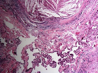
my first glimpse of the histology the next day:
Cholesterol
clefts and giant cells. Note in the upper picture that this
xanthogranulomatous reaction appears to be continuous with the choroid
plexus. Could this be a xanthogranuloma of the choroid plexus? I
considered that, until I looked elsewhere on the slide and saw this:
This is the ciliated epithelium characteristic of a colloid cyst of the
third ventricle, a tumor that can indeed "pop out" into the
neurosurgeon's hand. Gross inspection did not reveal a cyst
per se
because the exuberant xanthogranulomatous reaction obliterated it.
Cases like this have been described in the literature. For example, Dr.
David Louis and colleagues wrote in a 1994 article entitled
Third ventricle xanthogranulomas clinically and radiologically mimicking colloid cysts: Report of two cases
(Journal of Neurosurgery. 81(4):605-9, 1994 Oct.) that "histopathologic
examination revealed xanthogranulomas of the choroid plexus with only
microscopic foci of colloid cyst-like structures." I would argue quite
the opposite: that the xanthogranuloma derived from the colloid cyst and
extended into the choroid plexus. Indeed, Dr. Peter Burger and
colleagues write in the fourth edition of
Surgical Pathology of the Nervous System and Its Coverings that "a xanthogranulomatous reaction occasionally supervenes in colloid cysts, largely replacing the epithelium in some cases."
Cool case.


2 comments:
Excellent case Dr. Moore!! MAG.
Thanks a lot for sharing. Really excellent case.
Post a Comment