Monday, January 4, 2016

Best Post of July 2015: Medulloblastoma Markers

The next in our "Best of the Month" series is from July 17, 2015:

Four subgroups of medulloblastoma have been defined based on genetic alterations:

Wingless (WNT) - WNT medulloblastomas display monosomy 6 and most show nuclear accumulation of the WNT pathway protein beta-catenin, which serves as a useful immunohistochemical screen for this group. Medulloblasomas with more than 50% nuclear staining for beta-catenin have been shown to have WNT pathway activation, whereas those with only focal nuclear staining do not. Overall survival for WNT medulloblastomas are dramatically longer than those of other subtypes, and clinical practices surrounding the treatment of this subtype reflects this better prognosis.

Sonic Hedgehog (SHH) - SHH medulloblastomas often show a nodular/desmoplastic histopathology and are associated with a better prognosis in younger children and infants. 9q deletion is characteristic, and MYCN amplifications are occasionally noted. GAB1 is expressed in the cytoplasm of nearly all SHH medulloblastomas but not in other groups and can be detected imunohistochemically, making it a valuable SHH-group marker.  Targeted therapies directed at this subgroup have been established and are entering clinical practice.

"Group 3" - Group 3 medulloblastomas have the worst overall prognosis, have a high incidence of large cell/anaplastic histology, and are very frequently metastatic. This group contains the vast majority of MYC amplified tumors, with MYC amplification being a strong negative prognostic factor. It has been suggested that Group 3 tumors should perhaps be re-named MYC medulloblastomas, but wide agreement has not been reached on this designation. Group 3 tumors occur more commonly in males than females, and are found in infants and children, but almost never in adults.

"Group 4" - Group 4 medulloblastomas classically harbor isochromosome 17q; but as the molecular pathogenesis of this group is not currently clear, the generic name "Group 4" remains the consensus designation. Although isochromosome 17q is also seen in Group 3 tumors, it is much more common in Group 4. KCNA1 has been suggested as an immunohistochemical marker for this group, but this requires validation.  The only other notable cytogeneic change seen in Group 4 tumors is loss of X chromosome, which is seen in 80% of females with this tumor subtype. Group 4 patients have an intermediate prognosis, similar to patients with SHH tumors.

In conclusion, the worst prognosis is associated  with Group 3 medulloblastoma; Group 4 and SHH have an intermediate prognosis; and WNT medulloblastoma tends to have the best prognosis.

Saturday, December 19, 2015

Tuesday, December 15, 2015

Please show your gratitude to outgoing AANP Business Manager Peggy Harris

The Incomparable Peggy Harris
An important message from Mark Cohen, MD and Kathy Newell, MD:Peggy Harris needs no introduction to members of the American Association of Neuropathologists, as over the past 13 years she has supported and guided most of us (including the Executive Council and Committee Chairs) through the often confusing mechanics and byways of the society's functions, and has been instrumental in facilitating every aspect of our Annual Meetings, from abstract submission to registration to finding other members to making sure that everyone got CME credits. It would be almost impossible to list everything that Peggy has done for the AANP during her tenure with us. Pardon the neuropathological pun, but it would not be hyperbole to say that Peggy has been both our pia mater and glia during her tenure with the Association.

Peggy recently learned that she will no longer be assisting the AANP membership in the new year. Many of us will be sad to see her go, and will miss her at our Annual Meetings. For those who wish to contribute to a monetary gift of appreciation and support honoring Peggy's many contributions to the AANP, we have set up this special farewell fund.

This is not an official AANP-sponsored fundraiser. All proceeds donated to this fund will go directly to Peggy to show our gratitude to her. Please join us in showing how much we appreciate all that she has done for each of us individually and for the AANP as a whole. Thanks.

Friday, December 11, 2015

"I’m a neuropathologist with a glioblastoma": A guest post from Susan M Staugaitis, MD, PhD

I’m a neuropathologist with a glioblastoma.  How ironic is that?  For 17 years, my skill in making this diagnosis has been a major part of my job.  I know more about the biology of my disease than most of the physicians involved in my care.  And suddenly, within two weeks, my life expectancy changed from 30+ years to 2-5.  My reality has changed.

If I'm freaking you out, you are not alone.  I'm freaking everybody out.  I haven’t had such a good excuse to be so “frontal lobe” since I was a teenager.  In the past five months, I have become so accustomed to my diagnosis and disabilities that I blurt it out to store clerks when explaining why I’m seeking a particular good or service.  I know the song I want to hear over and over again as I cross to the other side.  Would you expect anything less from someone who looks a lot better than her scan?  

I cope by trying to stay realistic and in the moment.  I try to extract anything and everything that is positive from my experience today and defer to tomorrow all thoughts that won’t matter until then.  It’s not much different from the days when I lived in southern California, not worrying about the “big one” that could hit anytime, but keeping myself prepared nevertheless. 

Dr. Staugaitis wearing the Novocure Optune electric field generator as part of her treatment 
Family and friends remark on the extraordinary strength and courage I display.  Honestly, hearing this can get to be too much.  I do need the people close to me to help me fortify and reinforce the strength I am finding in myself, but quixotic sentiments don’t provide much comfort.  I’m too honest and practical to expect a miracle, to dream the impossible dream.  I’ll be satisfied with the possibility of beating the odds.

But, any gambler will tell you that the odds depend on the game.  I think sports gambling is most relevant to me and my diagnosis.  In sports gambling, it is all in the point spread.  You can win when your team loses and lose when your team wins.

Neuropathologists have inside information in this respect.  We look at the location of the lesion, at the histology in the microscope, at the molecular profile, and the combination gives us a pretty good idea of how to place our bet.  No emotions or wishful thinking here; we’ve got data.  As scientists, we are obligated to describe the data accurately and analyze it objectively.  In fact, if we are to gain and maintain the respect of our peers, we cannot ignore any data, no matter how much we dislike it, no matter how much it conflicts with our view of how the world should be.  And as writers, we find the words that communicate our interpretations as definitively and positively as possible without being incorrect.

Then it is time to sit back and watch the game, wait for the final score.  Did my bet beat the spread? 

And regardless of the game’s outcome, there will always be another game: new questions, new data to acquire.  So we pick ourselves up, dust ourselves off, and start all over again.

© 2015 Susan M. Staugaitis, M.D., Ph.D. 
Retired Consultant Staff, Departments of Pathology and Neurosciences, Cleveland Clinic
Adjunct Assistant Professor of Molecular Medicine

Cleveland Clinic Lerner College of Medicine of Case Western Reserve University

Thursday, December 10, 2015

Renowned neuropathologist Peter Burger set to retire at the end of this month

Peter C. Burger, MD
I just got word that the illustrious Johns Hopkins neuropathologist Peter C. Burger, MD is set to retire at the end of this month. In an email yesterday, the 74-year-old professor informed me that he will continue to work on various neuropathology projects, but will not be signing out cases after December 31. He writes that he is "ready for a new phase of life without the daily responsibility of cases". His sage consultations on difficult cases will be missed, but I am sure his impact will continue to be felt in the field through his valuable publications.

Wednesday, December 9, 2015

Not allowing kids to play football is "our moral duty": Bennet Omalu, MD

In a recent New York Times opinion piece, pathologist Bennet Omalu strongly endorsed a policy that children should not play football. Here are excerpts from his essay:

"We've known since 1964 that cigarette smoking is harmful to your health. We’ve known for more than 40 years that alcohol damages the developing brain of a child. We’ve known since the mid-70s that asbestos causes cancer and other serious diseases. Knowing what we know now, we do not smoke in enclosed public spaces like airplanes; we have passed laws to keep children from smoking or drinking alcohol; and we do not use asbestos as an industrial product.

"If a child who plays football is subjected to advanced radiological and neurocognitive studies during the season and several months after the season, there can be evidence of brain damage at the cellular level of brain functioning, even if there were no documented concussions or reported symptoms. If that child continues to play over many seasons, these cellular injuries accumulate to cause irreversible brain damage, which we know now by the name Chronic Traumatic Encephalopathy, or C.T.E., a disease that I first diagnosed in 2002."

It should be noted that the CTE brain bank at Boston University found evidence of CTE in the brains of 131 out of 165 athletes who played professional, college, or high school football. In other words, based on this sample size, the risk of getting CTE from playing football for a relatively extended period is higher than the risk of getting lung cancer from smoking.

Since we have a legal age at which individuals are allowed to smoke, drink alcohol, and drive, it stands to reason that engaging in an activity that is likely to cause brain damage should also only be engaged in by individuals old enough to make an informed decision, says Omalu. 

Thanks to Dr. Chad Vanderbilt for alerting me to this article.

Tuesday, November 24, 2015

The Mercado Brain Cutting Device launched at the University of Colorado

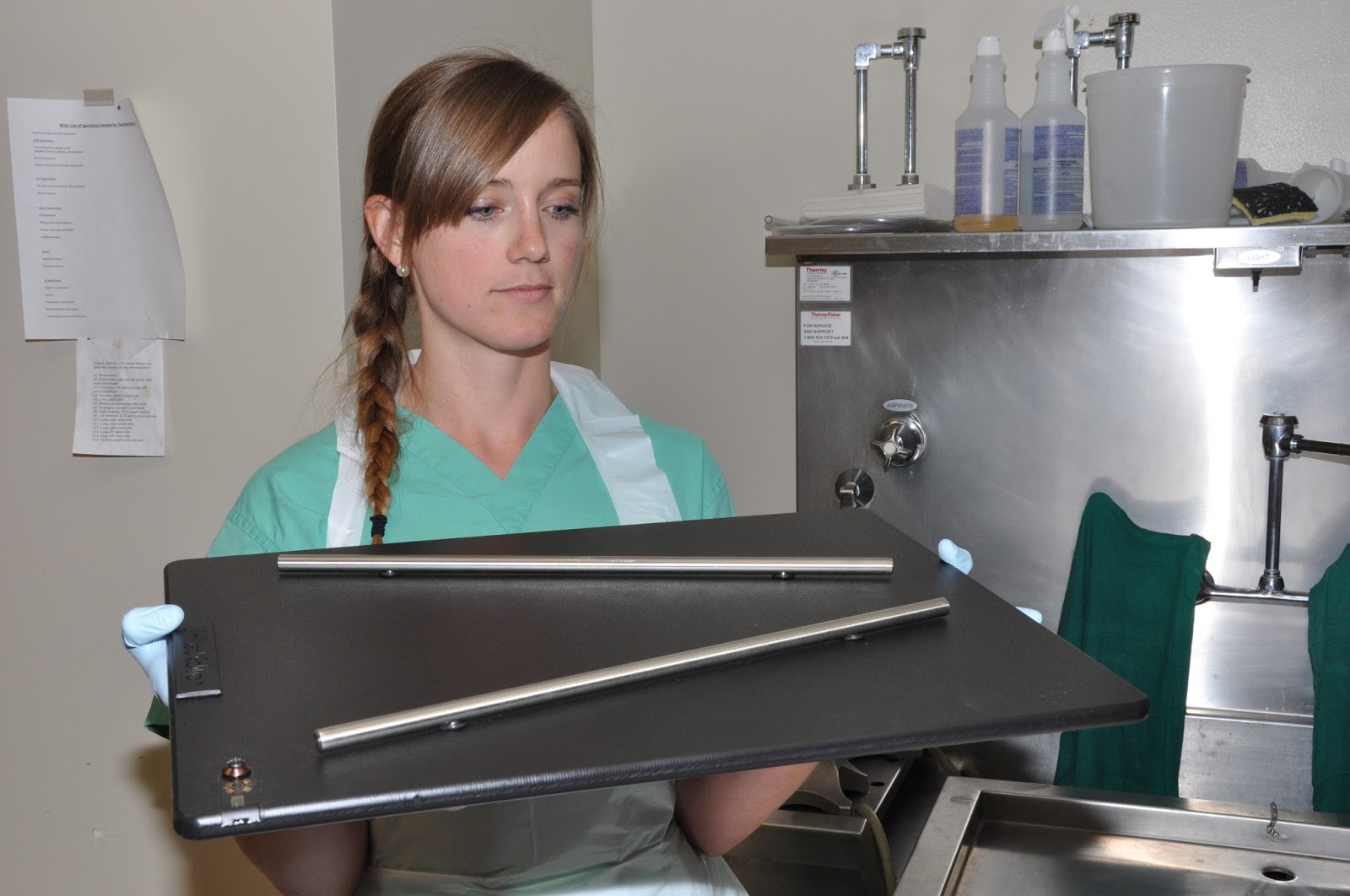
Thanks to a hand-made gift from University of Alabama neuropathology fellow Juan Mercado, MD, our residents on autopsy rotation this month had the opportunity yesterday to inaugurate the use of The Mercado Brain Cutting Device (MBCD). Made from tools easily found at any hardware store, the device allows prosectors to make reliably even 1-cm thick coronal brain slices for optimal demonstration of gross anatomy and pathology. University of Colorado pathology residents Abby Richmond, MD (PGY-III) and Sammie Roberts, MD (PGY-I) used the device to great advantage as demonstrated by the exquisitely presented brain slices laid out for inspection.

Dr. Sammie Roberts with the MBCD 
Dr. Abby Richmond makes the first cut

Sections are even and uniform in thickness

The finished product

Drs. Moore, Richmond, and Roberts (left to right) examining the coronal sections

Much appreciation to Dr. Mercado for gifting this device, which he describes as a "limited edition (1 or 1)", to our department. We will undoubtedly have more meticulous brain cutting sessions henceforth thanks to Dr. Mercado's efforts.

Wednesday, November 18, 2015

A 60-year-old woman with a left occipitotemporal brain lesion

We were sent this case in consultation to rule out infiltrating neoplasm. The diagnosis is amyloid beta related angiitis (ABRA). In this case, no infarction was present in the specimen, but white matter rarefaction (the presumed pathologic correlate to the leukoaraiosis reported on imaging) is present.

Granulomatous vasculitis associated with pink hyaline material in vessel walls

The pink hyaline material is shown to be beta-amyloid by immunohistochemistry

White matter rarefaction (upper half of picture) correlating with leukoaraiosis seen on imaging

Tuesday, November 17, 2015

Best Post of June 2015: First Day at 2015 American Association of Neuropathologists Meeting - The Special Course

The next in our "Best of the Month" series comes from 11 June 2015, where we harken back to the first day at the American Association of Neuropathologists (AANP) annual meeting held in my newly adopted home of Denver, Colorado:


The "Special Course" which typically launches the annual AANP meeting was anything but typical this year. Rather than the usual series of research presentations which has characterized the first day of the meeting, the 2015 edition of the "Special Course" focused on need-to-know practical topics. Dr. Beatriz Lopes put together a program that offered something for everyone -- from trainee to seasoned practitioner. The morning started with a presentation by Dr. Caterina Giannini on primary CNS lymphoma, as well as its mimickers and precursors. Far from being a straightforward diagnosis, lymphoma -- particularly in the setting of corticosteroid therapy -- can be mistaken for anything from multiple sclerosis to infarct. Next, two eminent scholars from Paris, Drs. Francoise Gray and Elisabeth Tournier-Lasserve, presented a three-part lecture on the hereditary non-amyloid small vessel diseases of the brain. Then Charles Eberhart showed up from Johns Hopkins to provide a primer on ophthalmic pathology, providing a practical approach to eye specimens. For example, Dr. Eberhart discussed the significance of uveal granulomatosis in orbital exenteration specimens. The presence of this finding should be noted in the pathology report as it portends an increased risk for sympathetic ophthalmia. Dr. Arie Perry appeared next with a presentation on the molecular characterization of brain tumors using immunohistochemical surrogates -- an approach particularly appreciated by neuropathologists who work at smaller institutions which may not have the resources to perform sequencing and other more advanced laboratory tests. After lunch, the focus was redirected to neuropathology training. Drs. Marc Del Bigio and Suzanne Powell discussed the state of neuropathology training abroad and in the United States, respectively. Finally, a lively discussion was initiated by a panel featuring Drs. Jeff Golden, Dennis Dickson, Liz Cochran, and myself organized under the title "The Professional Market for Neuropathology Trainees". Several audience members talked about their view of the profession and how its many facets are reflected in the training we give fellows and how that training impacts the preparedness of trainees for the job market. Overall, it was engaging day which, as audience member John Donahue put it, was "well worth the price of admission!"

Neuropathology Blog is Signing Off

Neuropathology Blog has run its course. It's been a fantastic experience authoring this blog over many years. The blog has been a source...