 |
| Screenshot of Dr. Perry's Lecture on YouTube |
Tuesday, April 24, 2018
YouTube has lecture by Dr. Arie Perry entitled "Approach to Diagnosis of Glial Neoplasms"
Looking for a good overview of glial neoplasms? There's a lecture on YouTube entitled Approach to Diagnosis of Glial Neoplasms by Dr. Arie Perry. Check it out!


Wednesday, April 18, 2018


Tuesday, April 17, 2018

Monday, April 16, 2018
Anaplastic Gangliocytoma in the frontal lobe of a 35-year-old female
This is the latest in several resections and follows two rounds of chemotherapy. Immunomarkers positive for neurofilament, and negative
for S100, GFAP, synaptophysin, chromogranin, NeuN, HMB45, and MelanA.
 |
| Anaplastic Gangliocytoma (note the necrosis in the lower right corner) |
Thursday, April 12, 2018
Colloid cyst of the third ventricle
 |
| Modified by compressive atrophy, the cyst lining cells would otherwise be columnar with obvious cilia |
 |
| A different area of the same cyst which more clearly demonstrates cilia |
Wednesday, April 11, 2018
There is a neuropathology museum in Lima, Peru
 The Institute of Neurological Science (INCN) in Lima houses the only "Brain Museum" in Latin America. The collection contains over 3000 samples from which around 300 are on permanent display, including brains with diverse cerebral diseases and anomalies as well as fetal brains with abnormalities. Unique photomicrographs and other items are also on display.
Monday, April 9, 2018
Posterior fossa tumor in a 5-month-old girl
The tumor was GAB1 positive (a marker for the SHH pathway).
 |
| Desmoplastic./nodular medulloblastoma |
Tuesday, April 3, 2018
Exuberant endothelial reactive hyperplasia next to a subacute infarct biopsied to rule out neoplasia
 |
| Endothelial hyperplasia next to an infarct, not to be confused with microvascular proliferation in a glioma |
Wednesday, March 21, 2018
Best Post of January 2018: Choroidal ganglioneuronal hamartoma in an NF1 patient
The next in our "Best of the Month" series is from January 30, 2018:
Thanks to Dr. Ahmed Gilani (pediatric pathology fellow at the University of Colorado) for providing me with slides of an enucleation specimen from a patient with Von Recklinghausen Neurofibromatosis (NF-1). The specimen exhibits a region of choroidal expansion with hamartomatous neuroglial tissue. Distributed throughout this choroidal expansion are non-pigmented ovoid bodies, which have a delicately laminated appearance reflecting the presence of concentric Schwann cell processes. One might conceive of these choroidal expansions as cousins of iridic Lisch nodules.
Thanks to Dr. Ahmed Gilani (pediatric pathology fellow at the University of Colorado) for providing me with slides of an enucleation specimen from a patient with Von Recklinghausen Neurofibromatosis (NF-1). The specimen exhibits a region of choroidal expansion with hamartomatous neuroglial tissue. Distributed throughout this choroidal expansion are non-pigmented ovoid bodies, which have a delicately laminated appearance reflecting the presence of concentric Schwann cell processes. One might conceive of these choroidal expansions as cousins of iridic Lisch nodules.
 |
| Choroidal expansion in an enucleation specimen from a child with NF1 |
 |
| Ganglion-like cells within the choroidal expansion |
 |
| Ovoid body within choroidal expansion |
Monday, March 19, 2018
Epigenomics to Enhance Tumor Classification
The Neuropathology Department at Heidelberg University Hospital led by Professor Andreas von Deimling, have developed a new computer-based method. “We hope that our new molecular classification method will help improve diagnostic accuracy in CNS tumors and, thus, also improve the chances for successful treatment,” said von Deimling.
The researchers analyzed specific DNA methylations. Different cell types exhibit characteristic patterns of DNA methylation which enable scientists to draw conclusions about a tumor’s cellular origin. "We have developed computer-based algorithms that reliably differentiate 82 types of CNS tumors based on their methylation patterns," said Professor David Capper, who is one of the four first authors of the study. “Particularly in tumors which we cannot easily assign to a diagnostic category based solely on microscopic examination, methylation analysis is often helpful to make a precise diagnosis. The analysis of approximately 2,800 reference tumor samples additionally made it possible to classify tumors into specific subgroups that are not yet included in the classifications that have been used so far.”
In order to test whether the method is suitable for use in clinical routine diagnostics, the scientists analyzed more than 1,100 additional tumor samples. In about twelve percent of the cases, they were able to correct the initial diagnosis using the methylation patterns. In almost all cases where it was possible, further molecular-diagnostic examinations showed that molecular classification characterized the tumors even better than the initial microscopic diagnosis.
“We are convinced that our new method is well suited to be used in the clinic,” said Stefan Pfister, one of the paper's authors. He added: “We have made our classification system available online in order to enable researchers to analyze their data at our platform.”
Reference: Capper, D., Jones, D. T. W., Sill, M., Hovestadt, V., Schrimpf, D., Sturm, D., … Pfister, S. M. (2018). DNA methylation-based classification of central nervous system tumours. Nature. https://doi.org/10.1038/nature26000
Wednesday, March 14, 2018
On Einstein's Birthday, We Take a Second Look at His Brain
On this date 139 years ago, Albert Einstein was born in Ulm, Germany. We take this occasion to republish a post from November 21, 2012 entitled: Photos reveal unique features of Einstein's cerebral cortex:
Photographs taken shortly after his death, but never before analysed in detail, have now revealed that Einstein’s brain had several unusual features, providing clues about the neural basis of his extraordinary mental abilities.
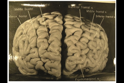
Nature.com reports that, while doing Einstein's autopsy, the pathologist Thomas Harvey removed the physicist's brain and preserved it in formalin. He then took dozens of black and white photographs of it before it was cut up into 240 blocks. Now, anthropologist Dean Falk of Florida State University in Tallahassee and her colleagues have obtained 12 of Harvey’s original photographs from the National Museum of Health and Medicine in Silver Spring, Maryland, analysed them and compared the patterns of convoluted ridges and furrows with those of 85 brains described in other studies.Many of the photographs were taken from unusual angles, and show structures that were not visible in photographs that have been analysed previously. The analysis was recently published today in the journal Brain. The most striking observation, says Falk, was “the complexity and pattern of convolutions on certain parts of Einstein's cerebral cortex”, especially in the prefrontal cortex, and also parietal lobes and visual cortex.
Photographs taken shortly after his death, but never before analysed in detail, have now revealed that Einstein’s brain had several unusual features, providing clues about the neural basis of his extraordinary mental abilities.
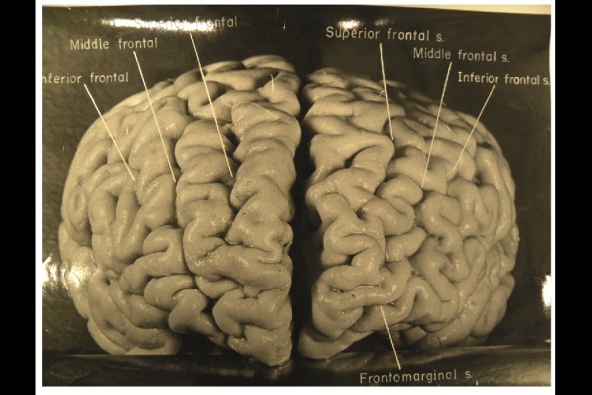
Nature.com reports that, while doing Einstein's autopsy, the pathologist Thomas Harvey removed the physicist's brain and preserved it in formalin. He then took dozens of black and white photographs of it before it was cut up into 240 blocks. Now, anthropologist Dean Falk of Florida State University in Tallahassee and her colleagues have obtained 12 of Harvey’s original photographs from the National Museum of Health and Medicine in Silver Spring, Maryland, analysed them and compared the patterns of convoluted ridges and furrows with those of 85 brains described in other studies.Many of the photographs were taken from unusual angles, and show structures that were not visible in photographs that have been analysed previously. The analysis was recently published today in the journal Brain. The most striking observation, says Falk, was “the complexity and pattern of convolutions on certain parts of Einstein's cerebral cortex”, especially in the prefrontal cortex, and also parietal lobes and visual cortex.
The autopsy revealed that Einstein’s brain was smaller than average and subsequent analyses showed all the changes that normally occur with ageing. Nothing more was analysed, however. Harvey stored the brain fragments in a formalin-filled jar in a cider box kept under a beer cooler in his office. Decades later, several researchers asked Harvey for some samples, and noticed some unusual features when analysing them.
A study done in 1985 showed that two parts of his brain contained an unusually large number of non-neuronal cells called glia for every neuron2. And one published more than a decade later showed that the parietal lobe lacks a furrow and a structure called the operculum3. The missing furrow may have enhanced the connections in this region, which is thought to be involved in visuo-spatial functions and mathematical skills.
AFP/Getty Images
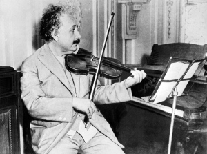
Einstein was a keen violinist, which may account for an overdeveloped section of his brain that deals with the left hand.
The prefrontal cortex is important for the kind of abstract thinking that Einstein would have needed for his famous thought experiments on the nature of space and time, such as imagining riding alongside a beam of light. The unusually complex pattern of convolutions there probably gave the region and unusually large surface area, which may have contributed to his remarkable abilities.
Falk and her colleagues also noticed an unusual feature in the right somatosensory cortex, which receives sensory information from the body. In this part of Einstein’s brain, the region corresponding to the left hand is expanded, and the researchers suggest that this may have contributed to his accomplished violin playing.
According to Sandra Witelson, a behavioural neuroscientist at McMaster University in Hamilton, Canada, who discovered that the parietal operculum is missing from Einstein’s brain, the study’s biggest contribution may be in encouraging further studies. “It makes clear the location and accessibility of photographs and slides of Einstein's brain,” she says. “This may serve as an incentive for other investigations of Einstein's brain, and ultimately of any consequences of its anatomical variations.”
Subscribe to:
Posts (Atom)
-
 Shannon Curran, MS with her dissection Shannon Curran, a graduate student in the Modern Human Anatomy Program at the University of Co...
-
 According to a recent Associated Press report , singer Michael Jackson's brain was removed from his skull and is being held for further ...