A group out of Wake Forest in North Carolina just published an article entitled Subconcussive Head Impact Exposure and White Matter Tract Changes over a Single Season of Youth Football in the journal Neuroradiology. Head impact data were recorded by using the Head Impact Telemetry system and quantified as the combined probability risk-weighted cumulative exposure (RWEcp).
Twenty-five male participants were evaluated for seasonal fractional anisotropy (FA) changes in specific white matter tracts.There were statistically significant linear relationships between RWEcp and decreased FA in certain white matter tracts. This study found a statistically significant relationship between head impact exposure and change of white matter FA value of in the
absence of a clinically diagnosed concussion. This research supports work by Ann McKee and others hinting at histologic changes that can be incidentally observed at autopsy among young football players. (Thanks for Dr. Adam King for alerting me to this important article from the radiology literature.)
Thursday, November 10, 2016


Tuesday, November 8, 2016
MOC Exam Topic: More on Aquaporin-4
My last post elicited two important comments on aquaporin-4. Since not all readers necessarily look at the comments, I am publishing them as a separate post here:
 Maria said...
Maria said...
Worth mentioning that [aquaporin-4] is the most well known target in Neuromyelitis Optica (NMO) and NMO spectrum disorders, since about 80% of patients with this syndrome will have circulating anti-aquaporin 4 antibodies. The IHC is useful when considering active NMOSD on a biopsy specimen by showing loss of staining (Neurology. 2015 Jan 13;84(2):148-58)
Agent86 said...
And one can only get so far without mentioning the glymphatic pathway..
Genetic knock-out of the gene encoding the astroglial water channel aquaporin-4, which is importantly involved in paravascular interstitial solute clearance, exacerbated glymphatic pathway dysfunction after TBI and promoted the development of neurofibrillary pathology and neurodegeneration in the post-traumatic brain. These findings suggest that chronic impairment of glymphatic pathway function after TBI may be a key factor that renders the post-traumatic brain vulnerable to tau aggregation and the onset of neurodegeneration.
Iliff JJ, Chen MJ, Plog BA, Zeppenfeld DM, Soltero M, Yang L, Singh I, Deane
R, Nedergaard M. Impairment of glymphatic pathway function promotes tau pathology
after traumatic brain injury. J Neurosci. 2014 Dec 3;34(49):16180-93.
Genetic knock-out of the gene encoding the astroglial water channel aquaporin-4, which is importantly involved in paravascular interstitial solute clearance, exacerbated glymphatic pathway dysfunction after TBI and promoted the development of neurofibrillary pathology and neurodegeneration in the post-traumatic brain. These findings suggest that chronic impairment of glymphatic pathway function after TBI may be a key factor that renders the post-traumatic brain vulnerable to tau aggregation and the onset of neurodegeneration.
Iliff JJ, Chen MJ, Plog BA, Zeppenfeld DM, Soltero M, Yang L, Singh I, Deane
R, Nedergaard M. Impairment of glymphatic pathway function promotes tau pathology
after traumatic brain injury. J Neurosci. 2014 Dec 3;34(49):16180-93.
Monday, November 7, 2016
MOC Exam Topic: Aquaporin-4
Among the topics for the neuropathology maintenance of certification examination is aquaporin-4 (AQP-4). An important regulatory molecule in the maintenance of the proper flow of water across the blood-brain barrier, AQP-4 is the major water channel expressed within CNS astrocytic foot processes. Water flux across AQP4 is bidirectional.
Wednesday, November 2, 2016
Neuropathology Maintenance of Certification Topics
For those taking the maintenance of certification (MOC) examination at some point in the next few years, the American Board of Pathology (ABP) has published topics that may be covered in the examination. The examination consists of 150 multiple-choice questions, 50 of which are required to be in the a category designated "general neuropathology I". The remaining 100 questions can be from various categories which the examinee chooses (general neuropathology II, degenerative I & II, developmental/pediatric/congenital I & II, neoplastic I & II, and neuromuscular I & II). The ABP provides an MOC examination study guide to help examinees prepare for the exam. The following list is copied from the study guide and includes possible topics in the mandatory general neuropathology 50-question module:
abnormal corticospinal tracts/pyramids
leptomeningeal opacifications
acute hemorrhagic leukoencephalopathy
leukodystrophies
anaplastic astrocytoma
motor cortex; smear prep
aquaporin-4
multicystic encephalopathy
astrocytomas; chemotherapeutic resistance
multiple system atrophy
axonal injury; IHC
myxopapillary ependymoma
borderzone hypoxic-ischemic damage
orbital plate fractures
cervical spinal cord tracts
Pick disease
CNS cysts
pituitary gland histology
CNS neoplasms; loss of heterozygosity
postmortem artifacts
CSF; metastatic tumors
primary angiitis of the CNS
deep (basal) nuclei; tracts
ragged red fibers
dermatomyositis
retinal hemorrhage
dysembryoplastic neuroepithelial tumor
retinoblastoma
fetal developmental; neuroanatomy
skeletal muscle ultrastructure
fibrillary astrocytoma
spinal cord anatomy
glioblastoma
status marmoratus
GM1 and GM2 gangliosidoses
Steele-Richardson-Olszewski syndrome
hereditary sensory-motor neuropathies
substantia nigra
Huntington disease
tauopathies
hypoxic injury
TORCH infections
infant developmental neuropathology; cerebellum
tract degeneration
infant spinal cord; sequence of myelination
trinucleotide repeat disorders
Lafora progressive myoclonic epilepsy
vascular malformations
abnormal corticospinal tracts/pyramids
leptomeningeal opacifications
acute hemorrhagic leukoencephalopathy
leukodystrophies
anaplastic astrocytoma
motor cortex; smear prep
aquaporin-4
multicystic encephalopathy
astrocytomas; chemotherapeutic resistance
multiple system atrophy
axonal injury; IHC
myxopapillary ependymoma
borderzone hypoxic-ischemic damage
orbital plate fractures
cervical spinal cord tracts
Pick disease
CNS cysts
pituitary gland histology
CNS neoplasms; loss of heterozygosity
postmortem artifacts
CSF; metastatic tumors
primary angiitis of the CNS
deep (basal) nuclei; tracts
ragged red fibers
dermatomyositis
retinal hemorrhage
dysembryoplastic neuroepithelial tumor
retinoblastoma
fetal developmental; neuroanatomy
skeletal muscle ultrastructure
fibrillary astrocytoma
spinal cord anatomy
glioblastoma
status marmoratus
GM1 and GM2 gangliosidoses
Steele-Richardson-Olszewski syndrome
hereditary sensory-motor neuropathies
substantia nigra
Huntington disease
tauopathies
hypoxic injury
TORCH infections
infant developmental neuropathology; cerebellum
tract degeneration
infant spinal cord; sequence of myelination
trinucleotide repeat disorders
Lafora progressive myoclonic epilepsy
vascular malformations
Tuesday, November 1, 2016
Registration for AANP meeting launches today

The countdown to the 93rd Annual Meeting of the American Association of Neuropathologists starts today with registration now officially open. The meeting will be held in Garden Grove, CA on June 8-11, 2017. Go to the AANP Website to register online!
Thursday, October 20, 2016
AANP's new website a great improvement
The American Association of Neuropathologists Website Committee was formed in 2014 and has worked diligently over the past two years to launch a new website this past June. The new website provides increased functionality for both site visitors (open access) and AANP members (with login required). AANP self-assessment modules will continue to be hosted through the previous webpage host, Dayspring, accessible at www.neuropath-education.org.
Highlights of the new website include:
- Jobs Board
- Enhanced members-only area with editable profile and searchable member directory
- Announcements & Upcoming Events sidebar menu
- Membership renewal and event registration capability
Of particular note is the heroic effort Dr. Doug Anthony has put in as chair of the website committee. He deserves a meritorious award from the AANP!
Highlights of the new website include:
- Jobs Board
- Enhanced members-only area with editable profile and searchable member directory
- Announcements & Upcoming Events sidebar menu
- Membership renewal and event registration capability
Of particular note is the heroic effort Dr. Doug Anthony has put in as chair of the website committee. He deserves a meritorious award from the AANP!
 |
| Douglas C. Anthony, MD, PhD |
Tuesday, October 18, 2016
Brain Cancer Surpasses Leukemia as #1 Pediatric Cancer Killer
The following post appeared on the Johns Hopkins Neuropathology Blog last month. The author is Andrew Black:
New data from the CDC shows the mortality rates for pediatric cancers is in decline. A study published by the CDC found that during 1999–2014, the cancer death rate for patients aged 1–19 years in the United States dropped 20%. What is also changing are the type of patients dying. In 1999, leukemia was the leading killer of childhood cancer. That has been replaced by brain cancer. Numerous other trends were also observed in the study.
In both 1999 and 2014, more than one half of all cancer deaths among children and adolescents 1-19 years old were attributable to either leukemia or brain cancer. 3 out of 10 cancer deaths among children and adolescents aged 1–19 years in 1999 were due to leukemia (29.7%), and 1 in 4 were due to brain cancer (23.7%). By 2014, these percentages reversed and brain cancer was the most common site, accounting for 29.9% of total cancer deaths.
Thursday, October 13, 2016
Constructing Comments in a Pathology Report: Advice for the Pathology Resident
In an editorial in the current issue of Archives of Pathology and Laboratory Medicine entitled Constructing Comments in a Pathology Report: Advice for the Pathology Resident, Drs. Stephen Smith and Martha Yearsley of Ohio State University provide important points to remember when crafting a surgical pathology report. Among the points the authors is one of my pet peeves: the inclusion of the statement "Clinical correlation is required." Well, OF COURSE CLINICAL CORRELATION IS REQUIRED! It is almost insulting to the clinician to state that he or she must integrate all pieces of data into the care of the patient. That is a doctor's job -- whether it be surgeon, internist, or pathologist! Here's what Drs. Smith and Yearsley have to say on the subject:
"The complex circuitry of many a pathologist's brain in the creation of pathology reports has, in many cases, reflexively routed diagnoses through a small subcortical box en route to signing out the report—a box requiring the addition of a controversial phrase: “Clinical correlation is recommended” (CCIR). The question of whether a pathologist should append this 4-word phrase is one of some depth; after all, is not the function of the pathologist to clinically correlate the specimen for evaluation? Indeed, pathology cannot be practiced in a vacuum, devoid of clinical information, lest the risk of diagnostic error become unacceptably high. So who should clinically correlate, and when?
"The answer to that question is the prudent pathologist. It is our responsibility to obtain clinical information before addressing the microscopy before us. In cases when no history is available, an effort should likely be made to contact the clinician, pending the diagnosis...
"Some cases, however, cannot avoid CCIR. Tissues exhibiting pathology with a nonspecific or undetermined etiology warrant a comment (eg, a skin biopsy exhibiting nonspecific dermal chronic inflammation). Often, it is preferred practice to augment the presentation of the differential diagnosis in these cases: 'Based on the clinical and histologic findings, a diagnosis of X is favored; however, the differential diagnosis includes…' Yet again, the prudent pathologist takes control and clinically correlates. Is CCIR needed in this context? Certainly not, given that the prudent clinician will then clinically correlate any pathologic findings presented in a report, understanding that the pathologist cannot be definitive. Indeed, perhaps CCIR is best reserved as a statement for saying 'I cannot interpret these histologic findings without directly examining the patient' or 'I do not have enough clinical information available to interpret the histologic findings before me.' Most often, this scenario arises when limited clinical history is available and nonspecific histologic findings are seen that would require an exceptional degree of assumption on the part of the examining pathologist to definitively interpret. Great caution should be taken in making such assumptions."
So, the phrase "clinical correlation is recommended" can be thought of as code for a certain degree of justified uncertainty. I don't believe that code should be used in a surgical pathology report. As in every aspect of life, telling it like it is makes the most sense.
Friday, October 7, 2016
Best Post of August 2016: The utility of TTF-1 immunohistochemistry in the diagnosis of sellar region masses
The next in our "Best of the Month" series is from August 1, 2016:
Posterior pituitary cells (pituicytes) manifest nuclear TTF-1 positivity. Anterior pituitary cells are negative. Three rare neurohypophyseal neoplasms which are derived from pituicytes maintain TTF-1 nuclear positivity: pituicytoma, spindle cell oncocytoma, and granular cell tumor. At times when the neuropathologist is trying to distinguish one of these neoplasms from more common sellar tumors -- such as pituitary adenoma, meningioma, and schwannoma -- TTF-1 can be helpful.
Posterior pituitary cells (pituicytes) manifest nuclear TTF-1 positivity. Anterior pituitary cells are negative. Three rare neurohypophyseal neoplasms which are derived from pituicytes maintain TTF-1 nuclear positivity: pituicytoma, spindle cell oncocytoma, and granular cell tumor. At times when the neuropathologist is trying to distinguish one of these neoplasms from more common sellar tumors -- such as pituitary adenoma, meningioma, and schwannoma -- TTF-1 can be helpful.
 |
| TTF-1 immunohistochemistry in normal neurohypophysis |
Wednesday, October 5, 2016
Guest Post from Howard Chang, MD, PhD: Unknown White Matter Disease
I am please to present a guest post from the illustrious Dr. Howard Chang of Michigan State University, who presents a perplexing case. Dr. Chang would be interested in reader comments. He writes: "I can use some help from our colleagues. Any advice
(from anyone) on where and how to proceed for additional studies will be very
much appreciated."



 |
| Dr. Howard Chang |
This is a case of a 12-year-old male with cerebral palsy, severe
developmental delay (level 1-2 years), and seizures (stable, no seizure
episodes since 2 years). He had progressive decline in neurological functions
following flu-like illness. He received IVIG and steroids for clinical
diagnosis of GBS-CIDP (18 months prior to death). Initially he showed some
improvement, but neurological functions continued to decline, with multiple
hospitalizations. MRI imaging studies (2 weeks prior to death) showed
extensive abnormal signal of the cerebral and spinal white matter. He was made
DNR. A general autopsy including brain and spinal cord was performed.
General Autopsy:
1. Atrophy of low
extremity muscles and apparent atrophy of muscles of hands.
2. Cushingoid
appearance with central obesity, skin striations, and adrenocortical atrophy
(likely due to steroid therapy).
Neuropathology Autopsy:
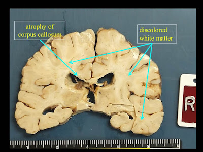
1. Extensive white matter atrophy-degeneration involving both the brain and spinal
cord (leukoencephalomyelopathy) with:
A. Microcephalic brain
(weight 1050 gm, normal should be about 1400 gm).
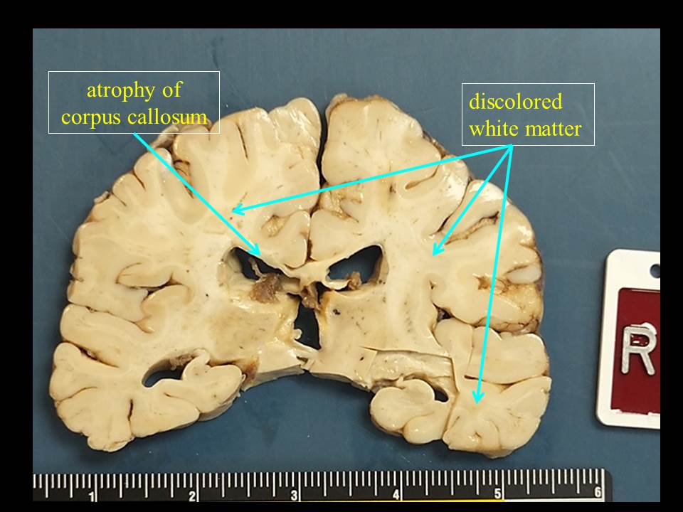
B. Bilateral cerebral
white matter atrophy-degeneration, with extensive astrogliosis and loss of
axons and myelin affecting the corpus callosum, and multifocal perivenous
microcystic changes involving the centrum semiovale, subcortical white matter,
with focal axonal spheroids in some of the microcystic areas.
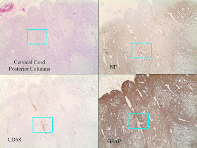
C. Spinal cord with
extensive microcystic degeneration of white matter tracts with loss of axons
and myelin, affecting bilateral posterior, anterior and lateral columns.
Focal loss of neurons within the spinal cord gray matter is noted, including
the anterior horn motor neurons and those in the Clarke’s nuclei. The nerve
roots appear relatively unremarkable.
D. Increased
perivascular macrophages are noted within the brain and spinal cord sections,
but there are no other areas of significant inflammation involving the brain or
spinal cord parenchyma, or the nerve roots. There is no obvious evidence
of abnormal cytoplasmic inclusions within either the neurons or glia.
2. Cerebral infarcts, small, involving the right occipital pole (subacute), and a
lacunar (old) infarct superior to the right occipital horn of the lateral
ventricle.


Monday, October 3, 2016
Mutant IDH1 and thrombosis in gliomas
 |
| Craig M. Horbinski, MD PhD |
Subscribe to:
Posts (Atom)
-
 Shannon Curran, MS with her dissection Shannon Curran, a graduate student in the Modern Human Anatomy Program at the University of Co...
 -
 Dr. PJ Cimino , whom we profiled when he was a fellow back in November of 2013, is a now faculty member at the University of Washington. I ...