 |
| Howard Chang, MD, PhD |
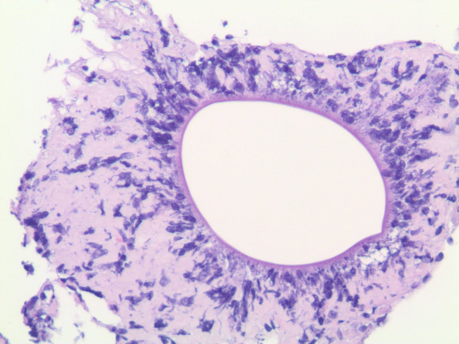
The illustrious
Dr. Howard Chang of Michigan State University recently sent me photomicrographs of a meningioma specimen from the posterior fossa of a 63-year-old female. Within the meningioma were noticed "funny worm-like profiles". The specimen was sent to the
Centers for Disease Control, which provided reassurance that these profiles were not parasites. The inimitable
Dr. John Donahue wondered whether this was simply embedded ependymal lining. I wondered the same thing. Dr. Chang tells me that GFAP was not ordered by the general pathologist signing out this case. Meanwhile, the eminent
Dr. Mark Cohen felt -- despite the CDC's pronouncement -- that the structures did not look human. Please help our colleague Dr. Chang by making any diagnostic suggestions in the comments section.


3 comments:
The unusual profiles are positive for CD34 and vimentin, but negative for desmin.
On behalf of Howard Chang:
I am not sure what these results indicate, but negative reactions for GFAP, S100, EMA, and pan-cytokeratin are features that argues against an epithelial or glial-ependymal differentiation. Negative reaction for desmin but positive reactions for vimentin and CD34 suggest mesenchymal but not muscle differentiation. Other thoughts?
As I have learned from previous lectures by very smart people, immunohistochemistry reactions color "what I don't know" brown... :(
Howard
After seeing similar profiles in the CUSA contents (and only in the CUSA) of several more meningiomas, I am concluding that these are most likely artifacts created by CUSA: Oil droplets created from cell membranes and other lipid membranes during sonication destruction of the cells (by CUSA), with adhering apoptotic nuclei, DNA, and other proteinacious debris on the outside of the oil droplets. Similar oil droplet artifacts may be seen in CUSA contents of other tumors, or hematoma.
Interesting insight, Howard. Thank you!
Post a Comment