Wednesday, December 7, 2016

Neuropathology History: Désiré Magloire Bourneville (1840-1909)

Désiré Magloire Bourneville (1840-1909)

Born in Garenciéres, France. From 1905 until his death, Bourneville headed the Foundation Vallée for the Study of Feebleminded Children.  "Was recognized as the leading continental authority on all aspects of mental abnormality of children. Most of his neuropathologic work was on idiocy. His description of tuberous sclerosis, since known as Bourneville's disease, appeared in 1880."

Source: Haymaker, Webb (Army Institute of Pathology). Guide to the exhibit on the history of neuropathology. Presented at the annual meetings of the American Psychiatric Association (Washington, DC, May 17-20, 1948) and the American Neurological Association (Atlantic City, June 14-17, 1948).

Tuesday, December 6, 2016

Neuropathology History: Sir Edward Farquhar Buzzard (1871-1945)

"Born in London... Became Regius Professor of Medicine at Oxford University, and consulting physician at St. Thomas's Hospital (1928). In the field of neuropathology, he is well known for his textbook in collaboration with Greenfield (1921)... Important also were his studies on myasthenia gravis (to which he contributed the term 'lymphorrhages') (1905), chronic progressive cerebral softening (1906),... delayed traumatic apoplexy (1909), and epidemic encephalitis (1919)."

Sir Edward Farquhar Buzzard (1871-1945)

Source: Haymaker, Webb (Army Institute of Pathology). Guide to the exhibit on the history of neuropathology. Presented at the annual meetings of the American Psychiatric Association (Washington, DC, May 17-20, 1948) and the American Neurological Association (Atlantic City, June 14-17, 1948).

Monday, December 5, 2016

Neuropathology History: Carl Wernicke (1848-1905)

"Born in Tarnowitz, Poland... A highly original thinker, he can be said to have been a pupil only of Meynert, though he was greatly influenced by the works of Hitzig and Munk."


Carl Wernicke (1848-1905)
Source: Haymaker, Webb (Army Institute of Pathology). Guide to the exhibit on the history of neuropathology. Presented at the annual meetings of the American Psychiatric Association (Washington, DC, May 17-20, 1948) and the American Neurological Association (Atlantic City, June 14-17, 1948).

Addendum: Comment from Dr. Darren Groh from Rhode Island Hospital: "Technically, Carl Wernicke was not born in Poland. He was born in the Kingdom of Prussia, which was part of Germany during his lifetime. This area is now part of Poland, however. I appreciate the history articles."

Friday, December 2, 2016

Guest Post: Fibrous Bodies Nicely Demonstrated in a Smear from a Somatotroph Pituitary Adenoma


Christian Davidson, MD

Dr. Christian Davidson, director of neuropathology at the Robert Wood Johnson University Hospital in New Jersey, provides today's blog post:

A 30-year-old man presented with bitemporal hemianopsia and a 3.0 cm pituitary mass was discovered upon MRI. His IGF-1 was elevated to 900, but he had no signs of acromegaly. A smear of tissue sent for frozen section evaluation (see below) revealed that most cells had round, eosinophilic, perinuclear inclusions suggestive of fibrous bodies (some examples are circled). Dot-like CAM5.2 immunostain (not shown) confirmed my smear-based diagnostic suspicion.


Thursday, December 1, 2016

Pineal Parenchymal Tumor of Intermediate Differentiation, WHO grade III


"A tumor of the pineal gland that is intermediate in malignancy between pineocytoma and pineoblastoma and is composed of diffuse sheets or large lobules of monomorphic round cells that appear more differentiated than those observed in pineoblastoma." -- WHO Book (2016)

The particular example depicted above recurred with leptomeningeal spread.

Tuesday, November 29, 2016

Vestige of a choroidal melanoma

Only melanin and melanophages remain in an enucleation specimen from a patient successfully treated with brachytherapy for choroidal melanoma. The eye was enucleated not because of the tumor, but because it was blind and intractably painful in the aftermath of treatment.


Monday, November 28, 2016

Best Post of October 2016: Brain Cancer Surpasses Leukemia as #1 Pediatric Cancer Killer

The next in our "Best of the Month" series comes from October 18, 2016:

The following post appeared on the Johns Hopkins Neuropathology Blog last month. The author is Andrew Black:

New data from the CDC shows the mortality rates for pediatric cancers is in decline. A study published by the CDC found that during 1999–2014, the cancer death rate for patients aged 1–19 years in the United States dropped 20%. What is also changing are the type of patients dying. In 1999, leukemia was the leading killer of childhood cancer. That has been replaced by brain cancer. Numerous other trends were also observed in the study.

In both 1999 and 2014, more than one ­half of all cancer deaths among children and adolescents 1­-19 years old were attributable to either leukemia or brain cancer. 3 out of 10 cancer deaths among children and adolescents aged 1–19 years in 1999 were due to leukemia (29.7%), and 1 in 4 were due to brain cancer (23.7%). By 2014, these percentages reversed and brain cancer was the most common site, accounting for 29.9% of total cancer deaths.

Wednesday, November 23, 2016

Germline and somatic BAP1 mutations in high-grade rhabdoid meningiomas

Ganesh Shankar of Brigham and Women's Hospital and colleagues recently published an article in Neuro-Oncology entitled Germline and somatic BAP1 mutations in high-grade rhabdoid meningiomas. Rhabdoid meningiomas are designated in the World Health Organization Classification of Tumours as high grade, despite the fact that only a subset follow an aggressive clinical course. To define genomic aberrations of rhabdoid meningiomas, the authors performed sequencing of cancer-related genes in 27 meningiomas from 18 patients with rhabdoid features and evaluated breast cancer [BRCA]1–associated protein 1 (BAP1) expression by immunohistochemistry in 336 meningiomas. The tumor suppressor gene BAP1 is inactivated in a subset of high-grade rhabdoid meningiomas. Patients with BAP1-negative rhabdoid meningiomas had reduced time to recurrence compared with patients with BAP1-retained rhabdoid meningiomas. A subset of patients with BAP1-deficient rhabdoid meningiomas harbored germline BAP1 mutations, indicating that rhabdoid meningiomas can be a harbinger of the BAP1 cancer predisposition syndrome. The authors conclude that BAP1-mutated rhabdoid meningiomas are clinically aggressive, requiring intensive clinical management.

Tuesday, November 22, 2016

MOC Exam Topic: Acute Hemorrhagic Leukoencephalopathy

First recognized as a discrete entity by Weston Hurst in 1941, acute hemorrhagic leukoencephalopathy (AHL) is a usually fatal disease characterized clinically by an abrupt onset of fever, neck stiffness, and neurological deficits, often progressing rapidly to seizures and coma. The presenting clinical picture is similar to that of acute disseminated encephalomyelitis (ADEM) but with a more fulminant course. At autopsy, the brain is swollen with multiple petechial hemorrhages centered in the white matter. Large foci of necrosis with cavitation may be present. The cerebral cortex and basal ganglia usually appear intact. Histologically, perivascular demyelinating lesions consist of ball or ring hemorrhages surrounding necrotic venules. There are cuffs of mononuclear cells and neutrophils. There is also substantial axonal injury in the affected areas. The lesions are indistinguishable from ADEM, but the extent of microvascular damage and therefore hemorrhage is is greater. An allergic mechanism is postulated.

FIGURE 3. A (H&E, 100×), B (LFB/PAS, 100×), and C (HAM-56 IHC, 400×). Light microscopic studies revealed thin sleeves of pallor surrounding small-caliber parenchymal blood vessels (A) which correspond to areas of demyelination on special stain (B). Macrophages stain strongly positive for macrophage marker HAM-56 (C). From Lann MA, et al.  2010 Mar;31(1):7-11.

Wednesday, November 16, 2016

Best Post of September 2016 - Featured Neuropathologist: Karra A. Jones, MD, PhD

The next in our "Best of the Month" Series is from September 6, 2016:
Karra Jones, MD, PhD
From time to time on Neuropathology Blog, we profile a prominent neuropathologist. In the past, we've featured the likes of Craig HorbinskiRoger McLendon, and Jan Leestma. Today, we feature a rising star in the field: Karra A. Jones, MD, PhD. Having just moved to the University of Iowa from UCSD, Dr. Jones is poised to do great work at her new institution. Here's a short bio followed by a Q&A with the inimitable Dr. Jones:

Karra Jones grew up in Kansas City where she completed her M.D. and Ph.D. at the University of Kansas School of Medicine. Karra’s graduate work focused on the evaluation of muscle spindle innervation by large peripheral nerve fibers and proprioceptive abnormalities in diabetes. During her time at KUMC, Karra was inspired by the strong history of neuropathology in Kansas City started by the dearly missed John Kepes and continued by her amazing mentor Kathy Newell. Karra traveled to the West Coast in 2010 to train in combined anatomic pathology/neuropathology under Lawrence Hansen, Scott VandenBerg, Subhojit Roy, and Henry Powell at the University of California, San Diego. There she focused on brain tumor research with Scott VandenBerg and Steve Gonias and developed a clinical interest in neuromuscular pathology. She was fortunate to obtain additional training in muscle pathology at UCSD with Diane Shelton in The Comparative Neuromuscular Laboratory. Karra joined the staff at UCSD in 2014 where she headed the neuromuscular service, participated in the general neurosurgical service, collaborated with molecular pathology on brain tumor molecular testing protocols/testing, supervised a biomarker laboratory, and was a co-director of the tissue biorepository. Karra very recently returned to the Midwest to join the highly talented neuropathology group at the University of Iowa where she is very excited to be practicing alongside Steve Moore, Leslie Bruch, Pat Kirby, and Gary Baumbach. 

1. Why did you decide to become a neuropathologist?

I became interested in the neurosciences after spending a year as a research assistant at Emory University in the Department of Neurology prior to medical school. Then, during graduate school at KUMC, my interest in tissue morphology was peaked after spending hours each day under a confocal microscope staring at muscle spindle innervation (what a gorgeous thing!) While at KUMC, I was extremely lucky to have Kathy Newell take me on as a mentee, and after that I was hooked. Almost everyone in my family is an artist, and I often felt like the outsider in that regard. But I realized with pathology, and in particular the beauty of neuropathology, I was a different kind of artist in my own right. Examining, classifying, and photographing brain tumors, neuromuscular diseases, and neurodegenerative diseases seemed like the most fun I could ever have at work. And I continue to have fun every day as a neuropathologist. 

2. Name a couple of important professional mentors. Why were they important to you?
I already mentioned Kathy Newell – Kathy has been an amazing mentor throughout my training and early career even though I haven’t worked with her directly since medical school. She first inspired me to pursue neuropathology with her amazing eye, calm demeanor, and kind heart. She also taught me about the importance of a “Neuropathology Family” introducing me to John Kepes and encouraging me to work with B.K. DeMasters during my last year of medical school, which helped solidify my decision to pursue combined AP/NP training. Another very important mentor is Lawrence Hansen (although he would argue that mentor means “cross-dresser” as the word is derived from Homer’s Odyssey in which Athena assumes the form of Mentor.) Larry is one of the most talented teachers and morphologists I have ever had the opportunity to work with. His “Hansen-isms” are embedded in my brain for life and as a neuropathologist and educator I will forever pass them on to my fellows, residents, students and mentees. Not only is Larry an amazing teacher and mentor, but also he is a very good friend and human being. I was also extremely lucky to be mentored by Scott VandenBerg on brain tumor diagnosis, molecular testing, and basic science research. Without Scott’s influence, I wouldn’t be where I am today. 

3. What advice would you give to a pathology resident interested in doing a neuropathology fellowship?

Do it! Neuropathology is clearly the best of all pathology specialties. But in all seriousness, Neuropathology training will give you a highly desirable skill set that will prepare you for a large variety of career paths. There are many ways to “differentiate” as a neuropathologist – academic, private practice, research, clinical, tumors, neuromuscular, neurodegenerative, etc. So, prior to your NP training, try to think about what you would like to do as a career after it’s all said and done, but remember to always be flexible and allow yourself to change your mind (it happens in medicine quite often). Neuropathology can also be a good specialty to combine with others such as pediatric pathology and forensic pathology, making you a highly desirable job candidate for varied positions. Don’t be intimidated by the 2 year commitment of the NP fellowship. One extra year in training is nothing in the grand scheme of life and only prepares you even better for the day you click “finalize” on your first case (or it gives you more time to work toward getting grant funding before the clock starts ticking). Right now, there are many job openings in neuropathology – we need bright, motivated, and enthusiastic trainees to become the next generation of neuropathologists. 


4. What city would you like a future American Association of Neuropathologists meeting to be held and why?


I would love for the meeting to be held in San Francisco again. I love visiting the city and always look for excuses to return.

Tuesday, November 15, 2016

MOC Exam Topic: Status Marmoratus

The neurons of the infant caudate, putamen, thalamus, and globus pallidus are susceptible to damage by hypoxia-ischemia. In some extensive injuries, a marked gliosis occurs and, if the brain is actively forming myelin in that region, there is hypermyelination of the area with aberrant myelination of astrocytic processes. There is frequently also neuronal loss and mineralization of residual neurons. The resulting white, firm, marbled-appearing lesion is called status mamoratus. Thought to occur if a hypoxic insult happens before the age of 6 to 9 months, status marmoratus has been associated with complicated parturitions and acute febrile illness during the first year of life. Lesions in the basal ganglia occurring after the period of active myelination exhibit only gliosis associated with neuronal loss. (Source: Greenfield's Neuropathology, 8th Edition)

status marmoratus involving thalamus and basal ganglia

Friday, November 11, 2016

Calcifying Pseudoneoplasm of the Neuroaxis (CAPNON)

Approximately 59 cases of CAPNON have been reported in the literature, A non-neoplastic entity that can be found in either an intra-axial or extra-axial location, the pathogenesis of CAPNON is unclear but a reactive process has been favored. The outcome is generally considered to be excellent, with gross total resection typically curative. This case is somewhat unique in that it harbors adipose tissue.

Foci of calcification and fat are present in this midline example


Nodules of basophilic calcification


The calcifications have a chondromyxoid appearance


Surgery was complicated as the lesion encased the anterior cerebral arteries


References:
Aiken AH, Akgun H, Tihan T, Barbaro N, Glastonbury C. Calcifying pseudoneoplasms of the neuroaxis: CT, MR Imaging, and Histologic Features. American Journal of Neuroradiology 30 (2009) 1256-1260.

Duque SG, Lopez DM, de Mendivil AO, Fernandez JD. Calcifying pseudoneoplasms of the neuroaxis: Report of four cases and review of the literature. Clinical Neurology and Neurosurgery 143 (2016) 116-120.

Thursday, November 10, 2016

White matter tract abnormalities seen in football players receiving subconcussive blows

A group out of Wake Forest in North Carolina just published an article entitled Subconcussive Head Impact Exposure and White Matter Tract Changes over a Single Season of Youth Football in the journal Neuroradiology. Head impact data were recorded by using the Head Impact Telemetry system and quantified as the combined probability risk-weighted cumulative exposure (RWEcp).
Twenty-five male participants were evaluated for seasonal fractional anisotropy (FA) changes in specific white matter tracts.There were statistically significant linear relationships between RWEcp and decreased FA in certain white matter tracts. This study found a statistically significant relationship between head impact exposure and change of white matter FA value of in the
absence of a clinically diagnosed concussion. This research supports work by Ann McKee and others hinting at histologic changes that can be incidentally observed at autopsy among young football players. (Thanks for Dr. Adam King for alerting me to this important article from the radiology literature.)


Tuesday, November 8, 2016

MOC Exam Topic: More on Aquaporin-4

My last post elicited two important comments on aquaporin-4. Since not all readers necessarily look at the comments, I am publishing them as a separate post here:

Blogger Maria said...
Worth mentioning that [aquaporin-4] is the most well known target in Neuromyelitis Optica (NMO) and NMO spectrum disorders, since about 80% of patients with this syndrome will have circulating anti-aquaporin 4 antibodies. The IHC is useful when considering active NMOSD on a biopsy specimen by showing loss of staining (Neurology. 2015 Jan 13;84(2):148-58)

Agent86 said...
And one can only get so far without mentioning the glymphatic pathway..

Genetic knock-out of the gene encoding the astroglial water channel aquaporin-4, which is importantly involved in paravascular interstitial solute clearance, exacerbated glymphatic pathway dysfunction after TBI and promoted the development of neurofibrillary pathology and neurodegeneration in the post-traumatic brain. These findings suggest that chronic impairment of glymphatic pathway function after TBI may be a key factor that renders the post-traumatic brain vulnerable to tau aggregation and the onset of neurodegeneration.

Iliff JJ, Chen MJ, Plog BA, Zeppenfeld DM, Soltero M, Yang L, Singh I, Deane
R, Nedergaard M. Impairment of glymphatic pathway function promotes tau pathology
after traumatic brain injury. J Neurosci. 2014 Dec 3;34(49):16180-93.

Monday, November 7, 2016

MOC Exam Topic: Aquaporin-4

Among the topics for the neuropathology maintenance of certification examination is aquaporin-4 (AQP-4). An important regulatory molecule in the maintenance of the proper flow of water across the blood-brain barrier, AQP-4 is the major water channel expressed within CNS astrocytic foot processes. Water flux across AQP4 is bidirectional.

Wednesday, November 2, 2016

Neuropathology Maintenance of Certification Topics

For those taking the maintenance of certification (MOC) examination at some point in the next few years, the American Board of Pathology (ABP) has published topics that may be covered in the examination. The examination consists of 150 multiple-choice questions, 50 of which are required to be in the a category designated "general neuropathology I". The remaining 100 questions can be from various categories which the examinee chooses (general neuropathology II, degenerative I & II, developmental/pediatric/congenital I & II, neoplastic I & II, and neuromuscular I & II). The ABP provides an MOC examination study guide to help examinees prepare for the exam. The following list is copied from the study guide and includes possible topics in the mandatory general neuropathology 50-question module:

 abnormal corticospinal tracts/pyramids
 leptomeningeal opacifications
 acute hemorrhagic leukoencephalopathy
 leukodystrophies
 anaplastic astrocytoma
 motor cortex; smear prep
 aquaporin-4
 multicystic encephalopathy
 astrocytomas; chemotherapeutic resistance
 multiple system atrophy
 axonal injury; IHC
 myxopapillary ependymoma
 borderzone hypoxic-ischemic damage
 orbital plate fractures
 cervical spinal cord tracts
 Pick disease
 CNS cysts
 pituitary gland histology
 CNS neoplasms; loss of heterozygosity
 postmortem artifacts
 CSF; metastatic tumors
 primary angiitis of the CNS
 deep (basal) nuclei; tracts
 ragged red fibers
 dermatomyositis
 retinal hemorrhage
 dysembryoplastic neuroepithelial tumor
 retinoblastoma
 fetal developmental; neuroanatomy
 skeletal muscle ultrastructure
 fibrillary astrocytoma
 spinal cord anatomy
 glioblastoma
 status marmoratus
 GM1 and GM2 gangliosidoses
 Steele-Richardson-Olszewski syndrome
 hereditary sensory-motor neuropathies
 substantia nigra
 Huntington disease
 tauopathies
 hypoxic injury
 TORCH infections
 infant developmental neuropathology; cerebellum
 tract degeneration
 infant spinal cord; sequence of myelination
 trinucleotide repeat disorders
 Lafora progressive myoclonic epilepsy
 vascular malformations

Tuesday, November 1, 2016

Registration for AANP meeting launches today


The countdown to the 93rd Annual Meeting of the American Association of Neuropathologists starts today with registration now officially open. The meeting will be held in Garden Grove, CA on June 8-11, 2017. Go to the AANP Website to register online!

Thursday, October 20, 2016

AANP's new website a great improvement

The American Association of Neuropathologists Website Committee was formed in 2014 and has worked diligently over the past two years to launch a new website this past June. The new website provides increased functionality for both site visitors (open access) and AANP members (with login required). AANP self-assessment modules will continue to be hosted through the previous webpage host, Dayspring, accessible at www.neuropath-education.org.

Highlights of the new website include:
- Jobs Board
- Enhanced members-only area with editable profile and searchable member directory
- Announcements & Upcoming Events sidebar menu
- Membership renewal and event registration capability

Of particular note is the heroic effort Dr. Doug Anthony has put in as chair of the website committee. He deserves a meritorious award from the AANP!

Douglas C. Anthony, MD, PhD


Tuesday, October 18, 2016

Brain Cancer Surpasses Leukemia as #1 Pediatric Cancer Killer

The following post appeared on the Johns Hopkins Neuropathology Blog last month. The author is Andrew Black:

New data from the CDC shows the mortality rates for pediatric cancers is in decline. A study published by the CDC found that during 1999–2014, the cancer death rate for patients aged 1–19 years in the United States dropped 20%. What is also changing are the type of patients dying. In 1999, leukemia was the leading killer of childhood cancer. That has been replaced by brain cancer. Numerous other trends were also observed in the study.

In both 1999 and 2014, more than one ­half of all cancer deaths among children and adolescents 1­-19 years old were attributable to either leukemia or brain cancer. 3 out of 10 cancer deaths among children and adolescents aged 1–19 years in 1999 were due to leukemia (29.7%), and 1 in 4 were due to brain cancer (23.7%). By 2014, these percentages reversed and brain cancer was the most common site, accounting for 29.9% of total cancer deaths.

Thursday, October 13, 2016

Constructing Comments in a Pathology Report: Advice for the Pathology Resident

In an editorial in the current issue of Archives of Pathology and Laboratory Medicine entitled Constructing Comments in a Pathology Report: Advice for the Pathology Resident, Drs. Stephen Smith and Martha Yearsley of Ohio State University provide important points to remember when crafting a surgical pathology report. Among the points the authors is one of my pet peeves: the inclusion of the statement "Clinical correlation is required." Well, OF COURSE CLINICAL CORRELATION IS REQUIRED! It is almost insulting to the clinician to state that he or she must integrate all pieces of data into the care of the patient. That is a doctor's job -- whether it be surgeon, internist, or pathologist! Here's what Drs. Smith and Yearsley have to say on the subject:

"The complex circuitry of many a pathologist's brain in the creation of pathology reports has, in many cases, reflexively routed diagnoses through a small subcortical box en route to signing out the report—a box requiring the addition of a controversial phrase: “Clinical correlation is recommended” (CCIR). The question of whether a pathologist should append this 4-word phrase is one of some depth; after all, is not the function of the pathologist to clinically correlate the specimen for evaluation? Indeed, pathology cannot be practiced in a vacuum, devoid of clinical information, lest the risk of diagnostic error become unacceptably high. So who should clinically correlate, and when?

"The answer to that question is the prudent pathologist. It is our responsibility to obtain clinical information before addressing the microscopy before us. In cases when no history is available, an effort should likely be made to contact the clinician, pending the diagnosis...

"Some cases, however, cannot avoid CCIR. Tissues exhibiting pathology with a nonspecific or undetermined etiology warrant a comment (eg, a skin biopsy exhibiting nonspecific dermal chronic inflammation). Often, it is preferred practice to augment the presentation of the differential diagnosis in these cases: 'Based on the clinical and histologic findings, a diagnosis of X is favored; however, the differential diagnosis includes…' Yet again, the prudent pathologist takes control and clinically correlates. Is CCIR needed in this context? Certainly not, given that the prudent clinician will then clinically correlate any pathologic findings presented in a report, understanding that the pathologist cannot be definitive. Indeed, perhaps CCIR is best reserved as a statement for saying 'I cannot interpret these histologic findings without directly examining the patient' or 'I do not have enough clinical information available to interpret the histologic findings before me.' Most often, this scenario arises when limited clinical history is available and nonspecific histologic findings are seen that would require an exceptional degree of assumption on the part of the examining pathologist to definitively interpret. Great caution should be taken in making such assumptions."

So, the phrase "clinical correlation is recommended" can be thought of as code for a certain degree of justified uncertainty. I don't believe that code should be used in a surgical pathology report. As in every aspect of life, telling it like it is makes the most sense.

Friday, October 7, 2016

Best Post of August 2016: The utility of TTF-1 immunohistochemistry in the diagnosis of sellar region masses

The next in our "Best of the Month" series is from August 1, 2016:

Posterior pituitary cells (pituicytes) manifest nuclear TTF-1 positivity. Anterior pituitary cells are negative. Three rare neurohypophyseal neoplasms which are derived from pituicytes maintain TTF-1 nuclear positivity: pituicytoma, spindle cell oncocytoma, and granular cell tumor. At times when the neuropathologist is trying to distinguish one of these neoplasms from more common sellar tumors -- such as pituitary adenoma, meningioma, and schwannoma -- TTF-1 can be helpful.

TTF-1 immunohistochemistry in normal neurohypophysis

Wednesday, October 5, 2016

Guest Post from Howard Chang, MD, PhD: Unknown White Matter Disease

I am please to present a guest post from the illustrious Dr. Howard Chang of Michigan State University, who presents a perplexing case. Dr. Chang would be interested in reader comments. He writes: "I can use some help from our colleagues.  Any advice (from anyone) on where and how to proceed for additional studies will be very much appreciated."

Dr. Howard Chang
This is a case of a 12-year-old male with cerebral palsy, severe developmental delay (level 1-2 years), and seizures (stable, no seizure episodes since 2 years). He had progressive decline in neurological functions following flu-like illness. He received IVIG and steroids for clinical diagnosis of GBS-CIDP (18 months prior to death). Initially he showed some improvement, but neurological functions continued to decline, with multiple hospitalizations.  MRI imaging studies (2 weeks prior to death) showed extensive abnormal signal of the cerebral and spinal white matter. He was made DNR. A general autopsy including brain and spinal cord was performed.

General Autopsy:
1. Atrophy of low extremity muscles and apparent atrophy of muscles of hands.
2. Cushingoid appearance with central obesity, skin striations, and adrenocortical atrophy (likely due to steroid therapy).

Neuropathology Autopsy:
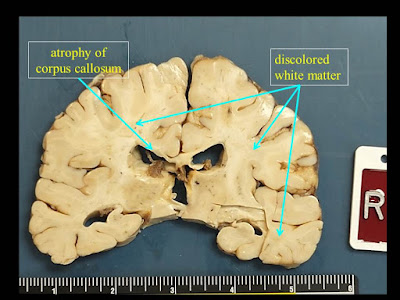
1. Extensive white matter atrophy-degeneration involving both the brain and spinal cord (leukoencephalomyelopathy) with:
A. Microcephalic brain (weight 1050 gm, normal should be about 1400 gm).

B. Bilateral cerebral white matter atrophy-degeneration, with extensive astrogliosis and loss of axons and myelin affecting the corpus callosum, and multifocal perivenous microcystic changes involving the centrum semiovale, subcortical white matter, with focal axonal spheroids in some of the microcystic areas.

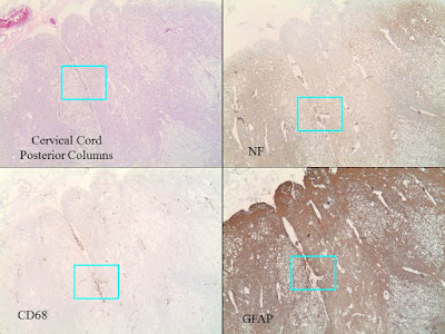
C. Spinal cord with extensive microcystic degeneration of white matter tracts with loss of axons and myelin, affecting bilateral posterior, anterior and lateral columns.  Focal loss of neurons within the spinal cord gray matter is noted, including the anterior horn motor neurons and those in the Clarke’s nuclei.  The nerve roots appear relatively unremarkable.

D. Increased perivascular macrophages are noted within the brain and spinal cord sections, but there are no other areas of significant inflammation involving the brain or spinal cord parenchyma, or the nerve roots.  There is no obvious evidence of abnormal cytoplasmic inclusions within either the neurons or glia.

2. Cerebral infarcts, small, involving the right occipital pole (subacute), and a lacunar (old) infarct superior to the right occipital horn of the lateral ventricle.


Monday, October 3, 2016

Mutant IDH1 and thrombosis in gliomas

Craig M. Horbinski, MD PhD
Dr. Craig Horbinski of Northwestern University and colleagues just published a significant paper in Acta Neuropathologica entitled Mutant IDH1 and thrombosis in gliomasHorbinski's data suggests that, quoting the abstract, "mutant IDH1 has potent antithrombotic activity within gliomas and throughout the peripheral circulation. These findings have implications for the pathologic evaluation of gliomas, the effect of altered isocitrate metabolism on tumor microenvironment, and risk assessment of glioma patients for venous thromboemboli." It should be noted that this paper won the Rubinstein Award at the 2015 American Association of Neuropathologists' meeting for Best Neuro-Oncology paper.

Thursday, September 29, 2016

Society for Neuro-Oncology meeting to feature several prominent neuropathologists


David W. Ellison, MD, PhD,
one of the presenters at the 2016 SNO meeting
The Society for Neuro-Oncology 2016 Annual Meeting to be held this year in Scottsdale, Arizona on November 16-20 will feature several members of the American Association of Neuropathologists. Peter Canoll will be speaking on "Primary brain tumors – pathology, grading and prognosis – new WHO classification". Arie Perry will be giving a "WHO overview". Sandro Santagata will talk on "Advances in the molecular genetics of meningioma". Dan Brat will present on "IDH-omas". Cynthia Hawkins will give an overview of pediatric gliomas. David Louis will give a keynote presentation entitled "The 2016 WHO classification of CNS tumors: an overview and a review of diffuse gliomas in adults". Additionally, Dr. Louis will be this year's recipient of the Victor Levin Award which honors an individual who has made a significant contribution to the field of neuro-oncology. David Ellison will provide a keynote presentation entitled "The 2016 WHO classification of CNS tumors - What’s new for pediatrics?" Finally, Fausto Rodriguez will give a talk entitled "Impact of NF1 status on glioma pathology". These individuals are advancing the field of neuropathology not only in their research but in their interactions with our colleagues in other fields of neuro-oncology including medical neuro-oncology, neurosurgery, neuroradiology, and radiation neuro-oncology.


Tuesday, September 27, 2016

Guest Post: Dr. Nat Pernick, Creator of PathologyOutlines.com

The following is a guest post from Dr. Nat Pernick, creator of PathologyOutlines.com, who reached out to me via Dr. Mark Cohen in search of neuropathology contributors to his website:


PathologyOutlines.com is looking for authors for its neuropathology chapters, specifically the CNS nontumor and CNS tumor chapters.  You can also write about Stains and Molecular Markers related to neuropathology.  You can either update existing topics or write about new topics.  More information is available on our Instructions for Authors page. The main advantages of being an author are: (a) you are helping the worldwide pathology community, as we average 15,000 daily visits; (b) it helps your academic career; and (c) and it may improve your writing skills.  We provide an honorarium of $20 per topic, but if money is your main motivation, this will not be a good fit.  You must be a good writer, and be able to follow our format.  You can write about one topic or many, but they must be approved in advance by us, and we have you do them one at a time.  Senior residents / fellows can write if they are supervised by a staff pathologist who meets our author requirements.  If interested, email your CV and a list of topics you want to write about (either update existing topics or write new topics) to Dr. Pernick at NatPernick@gmail.com.

Monday, September 26, 2016

University of Colorado residents show up in force for CAP16


Left to right: Drs. Robinson, Greer, Roberts, Klein, and Graham (with me kneeling)
Five PGY2 pathology residents, the most from a single class in the history of the institution, are presenting posters today and tomorrow at the annual meeting of the College of American Pathologists. Most are neuropathology related. Here are the titles:

Caleb Graham, et al. Pilocytic Astrocytoma: A Diagnostic Consideration in Lateral Ventricular Tumors

Ashley Greer, et al. BRAF Mutational Status in Desmoplastic Infantile Astrocytoma/Ganglioglioma

Colleen Klein, et al. Hemophagocytic Lymphohistiocytosis Discovered Clinically and at Autopsy: A Dual-Institutional, 10-Year Retrospective Review

Sammie Roberts, et al. Double Separate Versus Contiguous Pituitary Adenomas: Magnetic Resonance Imaging Features and Endocrinologic Follow-up

Chase Robinson, et al. Is Testing for IDH1 Mutation in Gliomas by Immunohistochemistry Worthwhile in Persons Older Than 55 Years?

Congratulations, everyone!

Sunday, September 25, 2016

CAP16 Scientific Plenary Session - The Immune Checkpoint Revolution in Cancer Treatment: "Moonshot" or "Pie in the Sky"

I'm attending the scientific plenary session at the 2016 annual meeting of the College of American Pathologists in Las Vegas. The discussion is being led by Dr. Lynette Sholl (pathologist), Dr. Christopher Lathan (medical oncologist), and Barry Nelson (lung cancer patient).  Although not directly related to neuropathology, the discussion of the role of biomarkers as a means of directing treatment impacts every area of surgical pathology -- including neuropathology. Mr. Nelson recounted his journey through immunotherapy. Dr. Sholl talked about the paradigm shift that has occurred in cancer therapy by virtue of harnessing T-cell mediated immunity, particularly related to PD-L1. Dr. Lathan, who is Mr. Nelson's physician, talked about his relationship with both the cancer patient and the pathologist. It was pointed out that everything starts with the diagnostic tissue; and the pathologist, as custodian of that tissue, is at the foundation of every patient's battle against cancer.  Importantly, Dr. Lathan pointed out that Mr. Nelson is a outlier. Most patients, at this point in our understanding of treatment, do not respond to immunotherapy. Finally, Mr. Nelson pointed out that he does not consider himself cured, but rather "healed". Thanks to immunotherapy, he lives with cancer rather than being cured from it. That relationship with cancer in itself is a paradigm shift, much like the one we have already gotten used to with regard to AIDS.

Left to right: Sholl, Nelson, Lathan

Thursday, September 22, 2016

A case of recurrent ligneous conjunctivitis in an adult

Massive fibrin deposition in a case of recurrent ligneous conjunctivitis in an adult

I recently signed out a case of ligneous conjunctivitis, a rare form of chronic pseudomembranous conjunctivitis that is marked by a massive accumulation of fibrin. The term ligneous (from the Latin term for "woody") refers to the firm consistency of the large masses of fibrin that comprise the pseudomembranes. Ligneous conjunctivitis typically occurs in children but may recur in adults. Treatment is often challenging because the inflammation is persistent and the pseudomembranes often recur rapidly after excision. Histopathology shows two components: granulation tissue and sheets of intensely eosinophilic acellular amorphous material, which has been shown to be composed predominantly of fibrin. The mass of fibrin also incorporates other serum components such as immunoglobulin. Lesions that resemble those found in the conjunctiva can affect other mucous membranes including the larynx, vagina, and ear. Ligneous conjunctivitis is an autosomal recessive trait caused by mutations in the gene for plasminogen on chromosome 6q26.

Reference: Eagle, Ralph C. Eye Pathology: An Atlas and Text [2nd edition] page 55.

Wednesday, September 21, 2016

CAP16 Abstract Highlights - BRAF Mutational Status in Desmoplastic Infantile Astrocytoma/Ganglioglioma

The 2016 annual meeting of the College of American Pathlologists (CAP16) is coming up September 25-28 in Las Vegas. In this series of posts, I'll be featuring poster abstracts of particular interest to neuropathologists.

Ashley Greer and colleagues at the University of Colorado in Poster #194 discuss the BRAF mutational status of DIA/DIG.


Context: Desmoplastic infantile astrocytoma/gangliogliomas (DIA/DIGs) are rare, massively cystic tumors usually found in superficial cerebral hemispheres. They are characterized by prominent desmoplastic stroma, interspersed neoplastic astrocytes, and fewer, if any, neoplastic ganglion cells. While BRAF mutation is found in up to 50% of pediatric gangliogliomas, 2 recent studies found it was rare in DIA/DIGs. We sought to assess BRAF mutation in our DIA/DIGs.

Design: Review of departmental files from 2000–2016 was performed to identify DIA/DIGs, with review of clinical outcome, neuroimaging features, immunohistochemistry (IHC) for astrocytic and neuronal markers, and BRAF VE1. BRAF mutational assessment was undertaken in IHC+/equivocal cases, with additional next generation sequencing whenever possible.

Results: All 6 cases were cerebral-hemispheric, with overlapping neuroimaging features (Figure 76, A and C) and favorable clinical outcomes, although histologic differences were noted. Five of 6 tumors contained a predominantly neoplastic astrocytic population (Figure 76, B), while DIA/DIG from the oldest child (12 months) showed exceptionally large nodules of neoplastic ganglion cells (Figure 76, D). This was the only case to show either BRAF VE1 IHC+ and/or mutation (rare c1799_1800TG.AT; p V600D). Next-generation sequencing on this case, and a comparison astrocytic-dominant DIA/DIG, showed only mutation in BRAF, and not in 26 other commonly mutated genes.

Conclusions: Five of 6 classic DIA/DIGs were negative for BRAF mutation. Previous publications found mutation in 2 of 18 and 1 of 14 cases; 2 of 3 reportedly mutated cases were unusual in that they were in atypical anatomic locations (suprasellar, fourth ventricle), and 1 was in an older child (24-month-old). DIA/DIGs with unusual features may be more likely to show BRAF mutation similar to ganglioglioma.

Tuesday, September 20, 2016

CAP16 Abstract Highlights - Astrocytoma With a Distinct Molecular Signature: MYB Rearrangement and EGFR Amplification

The 2016 annual meeting of the College of American Pathlologists (CAP16) is coming up September 25-28 in Las Vegas. In this series of posts, I'll be featuring poster abstracts of particular interest to neuropathologists.

Doan V. Lai and colleagues at Oklahoma University and St. Jude's in Memphis in Poster #184 describe a combination of low- and high-grade molecular features in a pediatric diffuse astrocytoma.

Diffuse gliomas are uncommon in children but cause significant morbidity and mortality. Unlike diffuse gliomas in adults, pediatric low-grade tumors rarely progress to high-grade disease. Molecular studies are playing an increasingly large role in classifying and predicting therapeutic response in these tumors. MYB rearrangements are common in pediatric diffuse low-grade astrocytomas. EGFR amplification, which occurs in high-grade gliomas, however, is not reported in low-grade gliomas. We report the first case of a pediatric astrocytoma with both MYB rearrangement and EGFR amplification. A 3-year-old boy was found to have a T2 hyperintense, nonenhancing mass in the
left temporal lobe after minor head trauma. Histology showed a diffuse astrocytic tumor with low cell density and bland cytology. Mitotic figures were present, albeit focally, prompting a diagnosis of anaplastic astrocytoma. No necrosis or microvascular proliferation was identified. The Ki-67 labeling index, while generally low, rose to approximately 20%, corresponding to regions of mitotic activity. Interphase fluorescence in situ hybridization analysis showed both rearrangement of MYB in two-thirds of cells and amplification of EGFR in approximately one-quarter of cells. This combination of low- and high-grade molecular features in a pediatric diffuse astrocytoma is so far unique and may
represent the molecular correlate of the rare clinical scenario where a pediatric diffuse astrocytoma, with MYB rearrangement, progresses to high-grade disease.

Saturday, September 17, 2016

CAP16 Abstract Highlights - Naegleria fowleri: Understanding the Clinical Presentation and Autopsy Findings of a Rare and Almost Universally Fatal Central Nervous System Infection

The 2016 annual meeting of the College of American Pathlologists (CAP16) is coming up September 25-28 in Las Vegas. In this series of posts, I'll be featuring poster abstracts of particular interest to neuropathologists.

Alexander T. Damron and colleagues at Baylor College of Medicine in Houston discuss Naegleria fowleri CNS infection in Poster #114:


Naegleria fowleri is a free-living ameba known to cause primary amebic meningoencephalitis (PAM). Moreover, PAM is an acute, fulminating, and hemorrhagic infection that occurs in healthy young children with fresh water exposure in warm climates. It is postulated that Naegleria fowleri enters through the nasal passages and crosses the cribriform plate, where it reaches the subarachnoid space and disseminates into the olfactory lobes. Visvesvara et al (2007) performed a retrospective study of all reported N fowleri infections in the United States from 1937 to 2013 and found 3 survivors in 142 reported cases. Only 27% of the 142 cases were diagnosed before patient death. We present a case of a previously healthy 14-year-old boy who presented with fever, headache, vomiting, and altered mental status 8 days after swimming in a warm freshwater lake. Cerebrospinal fluid studies showed organisms consistent with amoeba (Figure 265, C). Despite neuroprotective measures and antimicrobial medications, the patient was pronounced brain dead 9 days after admission. Autopsy revealed the cause of death to be PAM from infection with N fowleri with cerebral edema and tonsillar herniation (Figure 265, D). Microscopic examination of the central nervous system revealed amoebic organisms infiltrating the meninges and diffusely involving the brain parenchyma in a perivascular distribution (Figure 265, A and B). The most critical aspect in treating patients with PAM is early detection and prompt initiation of multiple antimicrobials and neuroprotective measures.Despite current recommendations, the high mortality rate of these infections (97%–98%) suggests that an effective treatment for PAM is not yet known.

Friday, September 16, 2016

CAP16 Abstract Highlights - Heterotopic Cutaneous Meningioma: An Unusual Presentation Occurring in a Patient With a History of Intracranial Meningioma

The 2016 annual meeting of the College of American Pathlologists (CAP16) is coming up September 25-28 in Las Vegas. I'll be attending the meeting this year. In this series of posts, I'll be featuring poster abstracts of particular interest to neuropathologists.

Chibuike L. Enwereuzo and Jean Henneberry at Baystate Medical Center/Tuft University School of Medicine, in Springfield, Massachusetts  discuss an unusual case of cutaneous meningioma in a patient with a history of intracranial meningioma (Poster #106):

Meningioma is a neoplasm of the meninges and typically occurs in intracranial sites. Extracranial meningioma has been reported most frequently in the sinonasal tract and skull bone, most often as extension of intracranial meningiomas. Isolated heterotopic meningioma without contiguous intracranial lesion is extremely rare. A 56-year-old woman presented in December 2015 with 2 firm subcutaneous scalp masses: one in the left lateral and the other in the left superior regions. The clinical impression was that of lipoma. Pathologic examination revealed
an ill-defined proliferation of fairly uniform meningothelial cells, infiltrating a fibroblastic stroma with interspersed adipose tissue. The 2 excised lesions had similar histologic features. Immunohistochemical stains were performed, and the tumor cells were positive for epithelial membrane antigen and negative for S100. A diagnosis of atypical meningioma, World Health Organization (WHO) grade II, was made. The patient’s medical history was significant for intracranial meningioma of the frontal lobe in October 2007, which was diagnosed as atypical meningioma, WHO grade II. Comparison of the scalp masses to her prior meningioma revealed a distinctly different morphologic pattern. Considering the 8-year interval between the scalp tumors and intracranial meningioma, it is unlikely that her most recent tumors represent a recurrence or metastasis of the intracranial tumor.

Dr. Diamandis develops network to help pathologists interface with AI computational scientists

A neuropathology colleague in Toronto (Dr. Phedias Diamandis) is developing some amazing AI-based tools for pathology and academia. He hel...