Friday, December 19, 2014

An inquiry for the neuropathology community from two illustrious German neurologists regarding possible olivary dysgenesis

Prof. Dr. Gunther Deuschl
Prof. Dr. Olaf Jansen


The 


The following is an inquiry received from Prof. Dr. Günther Deuschl, Chairman of the Department of Neurology, UKSH, Christian-Albrechts University, Kiel, Germany. Please post your ideas on this case in the comments section:

Dear friends,
I have a patient presenting with  different noises: one subjectively in the thyroid region (nothing objectively), one in the region of the ear (coming erratically in groups, some resemblance to earclicks, again nothing objectively), one pulse synchronous noise (with a known neuroradiologically confirmed and treated fistula carotis to sinus cavernosus). I would have considered her to have only the latter clinical problem. All the other complaints seemed to be due to enhanced introspection without definite psychiatric diagnosis. Then the 
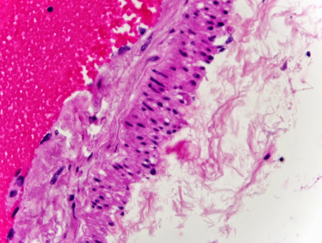
neuroradiologist (Prof. O. Jansen, Kiel University, Germany) made an MRI scan and found this curious ‘Tic-sign’ of the brain stem (you will understand when you look at the images below, which you can click on to enlarge). It shows definite circumscribed regional and bilateral atrophy of the inferior olive. Interestingly there seems to be some ‘white matter’ remaining. . Indeed an olivary agenesis or dysgenesis is one of the possibilities. The lady has no cerebellar signs as you see this sometimes (but not always) after olivary destruction.
I wonder if anybody has ever seen something like this and have an insights on a diagnosis?
Best regards for taking your time to read and see this.

Best regards,
Guenther


Tuesday, December 16, 2014

The Einstein/Rorke-Adams Connection

Dr. Lucy Rorke-Adams
I'm not sure how many in the neuropathology community know this, but our renowned colleague Lucy Rorke-Adams donated slices of Albert Einstein's brain to the Mütter Museum and Historical Medical Library in Philadelphia. Rorke-Adams, a senior neuropathologist at the Children's Hospital of Philadelphia, donated the 46 slices of brain tissue to the museum in 2011. Rorke-Adams had received the slides as a gift from local doctor, who in turn had bequeathed them from his colleague, a neuropathologist who examined the slides on behalf of Thomas Harvey, the man who removed Einstein's brain during an autopsy in 1955. Guess where my first stop will be on my next visit to Philly?

Monday, December 1, 2014

Best Post of September 2014: New ISN book on Peripherap Nerve Disorders coming soon

The next in our "Best of the Month Series is from September 26, 2014. The update on this post is that the book is NOW AVAILABLE!  The cost is a reasonable £119.11 (€148.80). (Don't ask me what that means in greenbacks!)

New ISN book – Peripheral Nerve Disorders – coming soon

A new book covering peripheral nerve diseases (part of the International Society of Neuropathology series)  is about to be released . The book editors are Jean-Michel Vallet and Joachim Weiss and includes chapters from a range of other international experts who have produced a clinically orientated guide to the pathology of peripheral nerve disorders. The book includes the latest molecular and pathological findings to provide the most up-to-date understanding of the pathogenesis of these disorders.

Tuesday, November 18, 2014

Robin Williams had Lewy Body Disease

Robin Williams
The official cause of Robin Williams' death, released Friday by the Marin County coroner, was ruled a suicide by hanging, with no evidence of alcohol or illegal drugs in his system and only therapeutic concentrations of prescribed medications.

Williams had long battled alcoholism, drug addiction and depression, but in November 2013 he was diagnosed with Parkinson's disease, according to his widow, after noticing a tremor in his left arm and difficulty moving on his left side as early as 2011.

Now a redacted pathology report from the autopsy on Williams' body has been made public and mentions "Diffuse Lewy body dementia,". Given that Dementia with Lewy Bodies can involve vivid visual hallucinations, it has now been speculated that perhaps such hallucinations may have lead to Williams' death.

Media reports, quoting anonymous "family sources," state that Williams' family believes that Lewy body disease was a critical "triggering" factor in his suicide. If so, this would be an unusual manifestation of the disease. Suicides have not been linked specifically to the hallucinations of Lewy Body Disease in the past.

Dennis Dickson, MD
"The use of the term dementia in the neuropathology report should not be inferred to mean that dementia was observed during life," warns Dennis Dickson of the Mayo Clinic in Jacksonville, who says he reviewed the neuropathology report.

"Mr. Williams was given a clinical diagnosis of (Parkinson's) and treated for motor symptoms. The report confirms he experienced depression, anxiety and paranoia, which may occur in either Parkinson's disease or dementia with Lewy bodies," Dickson said.

Thursday, October 30, 2014

"Atlas of Gross Neuropathology" to be released in the United States tomorrow!


I just received this nice email in my inbox this afternoon from a rising star in the neuropathology firmament. I hope that  readers will share their feedback on this new atlas in the comments section of Neuropathology Blog in the coming months:


Dear Brian:

I hope all is well for you.

I just wanted to let you know that our book is finally going to be released in the US tomorrow.


We would love to hear some feedback from people about the book. We hope it is useful in many settings, specialties and different levels of training and practice.

Very best wishes,


Sandra Camelo-Piragua, M.D.
Clinical Assistant Professor
Neuropathology Division
Pathology Department
University of Michigan

1301 Catherine St, MSB1  M4213
Ann Arbor, MI 48104
Phone: 734-936-1889
Fax:       734-615-2965

Friday, October 10, 2014

Best Post of August 2015: 35-year-old woman who died after a presumed seizure episode

The next in our "Best of the Month" series is from Thursday, August 21, 2014:

A 35-year-old woman died after a witness saw her "foaming at the mouth". A seizure was presumed to have occurred by emergency personnel. At autopsy, there was mild dilatation of the lateral and third ventricles:


This translucent mass was identified in the third ventricle:


Histological examination of the cyst contents revealed:

Although no ciliated epithelium was identified within the specimen, there were columnar cells with a pseudostratified configuration:


However, the majority of the cyst lining was flattened, simple epithelium:


Diagnosis is colloid cyst of the third ventricle. Normally one would see ciliated columnar cells lining the cyst, but they appear to be attenuated and flattened by the internal pressure of the cyst itself.

Thursday, October 2, 2014

My secret agent intercepted this communication

Editors: Seth Love, Arie Perry, James Ironside, Herbert Budka
Title: Greenfield's Neuropathology 9e
ISBN: 9781444166934
Publisher: Taylor & Francis
Estimated pub date: 20 February 2015
Estimated extent: 2000pp
Print run: 2000 copies

Listed on Amazon for only $778.35!!!!!!

Friday, September 26, 2014

New ISN book – Peripheral Nerve Disorders – coming soon

A new book covering peripheral nerve diseases (part of the International Society of Neuropathology series)  is about to be released . The book editors are Jean-Michel Vallet and Joachim Weiss and includes chapters from a range of other international experts who have produced a clinically orientated guide to the pathology of peripheral nerve disorders. The book includes the latest molecular and pathological findings to provide the most up-to-date understanding of the pathogenesis of these disorders.

Wednesday, September 24, 2014

Best Post of July 2014: 3D print of white matter "captures a sense of delicate complexity that evokes a sense of wonder about the brain"

The next in our "Best of the Month" series is from July 2, 2014:

Creating an accurate 3D model of the brain's white matter for Philadelphia's Franklin Institute was a project no 3D printing company would tackle -- until 3D Systems (Rock Hill, SC) agreed to take it on. Here is an image of the finished project, which took about 210 hours to print out:

Interviewed for the tech website CNET, Franklin Institute chief bioscientist and lead exhibit developer Dr Jayatri Das said that the model "has really become one of the iconic pieces of the exhibit. Its sheer aesthetic beauty takes your breath away and transforms the exhibit space," said . "The fact that it comes from real data adds a level of authenticity to the science that we are presenting. But even if you don't quite understand what it shows, it captures a sense of delicate complexity that evokes a sense of wonder about the brain."

Thanks to the illustrious Dr. Doug Shevlin for informing me of this remarkable feat of engineering which, in his words, sits at "the intersection of neuroscience, computers and 3D printing".

Friday, September 12, 2014

PBS's Frontline Spotlights Neuropathology in Documentary Film

In the film, which airs Tuesday, Oct. 8 beginning at 9 pm EST, FRONTLINE investigates the hidden story of the NFL’s response to head injuries. Through interviews with former players, scientists and neuropatholgists, it examines what the NFL knew about the risks of chronic traumatic encephalopathy (CTE), and when it knew it. Dr. Bennet Omalu, the first neuropathologist to draw a connections between playing football and the development of CTE, is featured in this remarkable documentary.

See a six-minute preview of the film here.

Dr. Bennet Omalu, the first pathologist to posit the connection between CTE and playing football

Thanks to Dr. Doug Shevlin sending me this link.

Tuesday, September 9, 2014

Amyloid-beta and neurofilament in 3D

Three-dimensional immunohistological visualization of Aβ (green) and neurofilament (red) in a 500-micron thick block of human post-mortem Alzheimer disease tissue.
 
From:Kunie A, et al. Inside Alzheimer brain with CLARITY: senile plaques, neurofibrillary tangles and axons in 3D. Acta Neuropathologica. , Volume 128, Issue 3, pp 457-459.

Thanks to Dr. Mark Cohen of Case Western for sending me the link.

Monday, September 8, 2014

Best Post of June 2014: Higgins Leads Athena's Quest for More Specific Epilepsy Diagnoses

The next in our "Best of the Month" series is from Thursday, June 5, 2014:

Joseph J. Higgins, MD
Last month I had an opportunity to sit down with Joseph J. Higgins, MD at the American Academy of Neurology Annual Meeting in Philadelphia. Dr. Higgins is Athena Diagnostics' recently named medical director for neurology.
Among the topics we discussed is Athena's new advances in helping clinicians diagnose autoimmune epilepsy disorders. A growing body of evidence points to an autoimmune etiology for a proportion of drug-resistant epilepsy cases. Dr. Higgins, with the horsepower of Athena's parent company Quest Diagnostics behind him, is leading the way in making auto-antibody testing available to patients with intractable epilepsy. While patients with pathogenic auto-antibodies often experience heightened adverse effects and typically respond poorly to conventional treatment, they can respond very well to immunomodulatory therapy. Autoantibodies to surface proteins that influence neuronal excitability have been found in the serum and cerebral spinal fluid of well over 10% of patients with epilepsy—whether the epilepsy was newly-diagnosed or established. In many cases, once identified, autoimmune epilepsy can be slowed, halted, or even reversed with adjunctive immunotherapy. Testing to establish an accurate diagnosis is an important part of selecting an optimal treatment plan. In a recent study (see reference below), 81% of adult patients diagnosed with autoimmune epilepsy experienced significant improvement in seizure status when immunotherapy was used in combination with anti-epileptic medications; 67% achieved complete seizure freedom.

Among the clinical syndromes that can be investigated using CSF auto-antibody assays are Morvan's Syndrome (by testing for VGKC complex, predominantly CASPR2) and NMDA Receptor Antibody Encephalitis (by testing for NMDA receptor, NR1 subunit). Dr. Higgins is at the forefront of personalized epilepsy treatment. Athena's advances in this field will ultimately improve patient outcomes while reducing medical costs. 


Reference: Quek AM, Britton JW, McKeon A, et al. Autoimmune epilepsy: clinical characteristics and response to immunotherapy. Arch Neurol 2012;69:582-93.

Monday, August 25, 2014

Best Post of May 2014: Primary Progressive Aphasia (non-fluent/agrammatic variant) in a patient with Pick disease

The next in our "Best of the Month" series is from May 14, 2014:

I recently performed an autopsy on a 67-year-old man had a seven-year history of progressive difficulty with halting speech. His wife described him as seeming to be “groping for words”. Three years after initial presentation, he demonstrated profound difficulty both initiating and finishing sentences. His verbal communication was marked by jumbled grammatical errors in which he put words in the wrong order and tense. For example, when asked by his neurologist to recount his activities over the day, he responded: “Go I… the grocery store… to.” He had no difficulty with naming objects or understanding their use. He had only mild amnestic deficits and no visuospatial difficulties. During the last months of his life, he began to become more rigid in his personality and more socially withdrawn. 
 
The clinical diagnosis in this patient is primary progressive aphasia, non-fluent/agrammatic variant. The pathologic diagnosis is frontotemporal lobar degeneration with 3R tau inclusions (FTLD-tau), commonly known as Pick disease.  The clinical presentation of FTLD can be divided into two types: the behavioral variant (bvFTLD) and primary progressive aphasia (PPA). Neurologists subclassify PPA into three subtypes: semantic variant (sv-PPA), non-fluent/agrammatic variant (na-PPA), and logopenic/phonologic variant (lv-PPA). While sv-PPA and na-PPA generally correspond to FTLD pathology, lv-PPA typically corresponds to Alzheimer disease (AD) pathology (and is accordingly referred to as the “language variant” of AD).  The syndrome of na-PPA is characterized by predominant language dysfunction in which there is a disordering of words in sentences, an inability to repeat complex words (e.g., saying “castaphory” when asked to repeat back the word “catastrophe”), inability to construct complex sentences, and other deficits in grammar and phonology.  In contrast, a diagnosis of sv-PPA is given to patients who exhibit a general deficit in their ability to understand word and object meaning. Patients with sv-PPA also show difficulty in naming objects and understanding what they are used for. A diagnosis of bvFTLD is ruled out as this patient only showed behavioral changes only late in the disease course

Pick disease was demonstrated pathologically by the presence of 3R tau positive Pick bodies (see photomicrograph) in the frontal and temporal lobes as well as the dentate fascia of the hippocampus, cornu Ammonis of the hippocampus, presubiculum, cingulate gyrus, insula, and inferior parietal lobe.

3R tau positive Pick body in the inferior temporal gyrus


For the practicing neuropathologist, FTLD’s are not yet classified on a molecular basis (although some day they surely will be),  but rather according to the particular protein aggregation which is detected using immunohistochemistry. The five possible FTLD subtypes, based on specific associated protein aggregations, are represented in the following table: 
FTLD type
Associated Protein Aggregation
FTLD-tau
Tau (can be further subclassified into 3-repeat [3R], 4-repeat [4R], or 3R+4R tau)
FTLD-TDP
Transactive response DNA-binding protein of 43 kDa (TDP-43)
FTLD-FUS
Fused in sarcoma (FUS)
FTLD-UPS
Ubiquitin (with UPS standing for ubiquitin-proteasome system)
FTLD-ni
No immunoreactivity
 
In 80% of cases of sv-PPA, the underlying histopathology is that of FTLD-TDP; while in the majority of na-PPA cases, the pathologic substrate is FTLD-tau.  Although not denoting a specific clinical syndrome, the term “Pick disease” has come to denote FTLD-tau, particularly referring to aggregation of the tau isoform in which there are three repeats (3R) of the phosphate-binding domain.

References:

Sieben A, Van Langenhove T, Engelborghs S, et al. The genetics and neuropathology of frontotemporal lobar degeneration. Acta Neuropathol. 2012;124(3):353-372.


Zdenek R, Matej R. Current Concepts in the Classification and Diagnosis of Frontotemporal Lobar Degenerations: A Practical Approach. Arch Pathol Lab Med. 2014;138:132-138.

Thursday, August 21, 2014

35-year-old woman who died after a presumed seizure episode

A 35-year-old woman died after a witness saw her "foaming at the mouth". A seizure was presumed to have occurred by emergency personnel. At autopsy, there was mild dilatation of the lateral and third ventricles:


This translucent mass was identified in the third ventricle:


Histological examination of the cyst contents revealed:


Although no ciliated epithelium was identified within the specimen, there were columnar cells with a pseudostratified configuration:


However, the majority of the cyst lining was flattened, simple epithelium:


Diagnosis?  (Please post to comments)


Tuesday, August 19, 2014

Dr. Peter Cummings Challenges Neuropathologists to Ice Bucket Challenge

Peter Cummings, MD
Dr. Peter Cummings has challenged his neuropathology colleagues to take the ALS ice bucket challenge. The good doctor dumps a bucket of ice water on his head in this video. If you are unfamiliar with the viral phenomenon of the ice bucket challenge, here's a New York Times article describing it. Now, grab a bucket!

Monday, August 18, 2014

Best Post of April 2014: Nobel Laureate Prusiner Tells His Story

The next in our  "Best of the Month" series is from April 8, 2014:

Just published by Yale University Press: Stanley Prusiner's new book, Madness and Memory: The Discovery of Prions - A New Biological Principle of Disease, is now available for purchase. Although I am not personally a big fan of Dr. Oliver Sacks's work, his blurb on Prusiner's book is worth reading: “Stanley Prusiner is a brilliant scientist whose boldness and tenacity enabled him, against all odds and despite near-universal skepticism, to discover and prove the importance of a new class of disease-producing agents—prions—a discovery as fundamental as that of bacteria and viruses. Prions, by subverting the brain’s own proteins, may play a crucial role in Alzheimer’s, Parkinson’s, and other neurodegenerative diseases—and perhaps afford a clue to their prevention. Madness and Memory is the story of one of the most important discoveries in recent medical history, and it is also a vivid and compelling portrait of a life in science." Special thanks to Dr. Mark Cohen for alerting me to the publication of this new account of a seminal discovery in biological science.

The bookish Dr. Mark Cohen

Monday, August 11, 2014

Best of March 2014: Vulnerability of Glioblastoma Cells to Catastrophic Vacuolization and Death Induced by a Small Molecule

The next in our "Best of the Month" series is from March 21, 2014:

Researchers at the Karolinska Institute in Sweden have introduced what could possibly be a revolution in glioblasoma treatment. In a recent article in Cell, researchers found that molecules known as vacquinols "reliably and selectively compromised" neoplastic cell viability. Vacquinols stimulate cell death by membrane ruffling, vacuolization, and -- ultimately -- cytoplasmic membrane rupture. Although in vivo testing has been restricted to mice thus far, this paper may prove to be the beginning of a new avenue of research into the selective killing of glioblastoma cells in patients.

Monday, August 4, 2014

Best Post of February 2014: Seizing Control of Brain Seizures

The next in our Best of the Month series comes from February 27, 2014:

How can trauma lead to chronic seizures? Berkeley researcher Daniela Kaufer found that only when albumin in the blood breaches the blood-brain barrier does the likelihood of post-traumatic epilepsy go up. Accelerated signaling between neurons results from this exposure, leaduing to seizures. “We were surprised, even a little disappointed, that it was such a common component of the blood  – nothing exotic at all  – that led to epilepsy,” recalls Kaufer, associate professor of integrative biology. She and colleagues went on to

Daniela Kaufer in the lab
show that albumin interacts with a ubiquitous cell protein TGF-Beta receptor to cause the damage. In the healthy brain, TGF-Beta signaling affects activity of astrocytes, which normally limit neuron-to-neuron firing signals across the synapse. But when albumin stimulates TGF-Beta receptors, astrocytes lose some of their control. Neuron signaling then spike dangerously, and promote the development of epileptic seizures. As luck would have it, statin drugs block TGF-Beta signaling.  Kaufer is now carrying out research to confirm that blocking abnormal TGF-Beta activity can prevent epilepsy from a range of insults. “Right now, if someone comes to the emergency room with traumatic brain injury, they have a 10 to 50 percent chance of developing epilepsy. But you don’t know which ones, nor do you have a way of preventing it. And epilepsy from brain injuries is the type most unresponsive to drugs." says Kaufer. “I’m very hopeful and that our research can spare these patients the added trauma of epilepsy.”

(Thanks to Dr. Doug "Scout" Shevlin for alerting me to this potentially groundbreaking research.)

Friday, July 25, 2014

Best Post of January 2014 -- The Alzheimer Conundrum: New Book Challenges Long-Held Assumptions

Margaret Lock, PhD
The next in our Best of the Month series comes from January 16, 2014: 
Professor Margaret Lock supplies an ethnographic account of Alzheimer disease in her recent book, The Alzheimer Conundrum. Lock furnishes a comprehensive description of the events leading up to the recasting of the phenomenon of Alzheimer’s as a condition to be prevented. The author challenges traditional assumptions and statistics about Alzheimer’s and takes us on a journey from the disease’s original clinical case through the vacillations in the science world and the media regarding possible causes, diagnostics, biomarkers, genetics and cures. She questions prevalence estimates for the impending “Alzheimer’s epidemic” that has been forecast with great assurance by some interest groups. Lock raises provocative questions that extend beyond the realm of Alzheimer disease, such as: When is a disease not a disease? When is pathology normal? When does a natural entity become pathological? Worth a read for those of us who are charged with the responsibility of ultimately diagnosing Alzheimer disease at autopsy.

Wednesday, July 16, 2014

"In a decade, the only people who are still playing football will be African-Americans and working class people." - Sociologist Harry Edwards

Harry Edwards, PhD
The public health policy debate (in which neuropathologists are inevitably enmeshed by virtue their involvement in the diagnosis of chronic traumatic encephalopathy) has taken on a new socio-economic ramification. I was reading an article this morning about NFL-player DeSean Jackson in the July 7th issue of ESPN The Magazine when I came across a provocative insight from sociologist Harry Edwards based on recent studies showing that white families and wealthier families are more informed about football-related concussions than their non-white and poor counterparts. Edwards had this to say about the changing demographics of football: "In a decade, the only people who are still playing football will be African-Americans and working class people."  Just as boxing was once embraced by Ivy League athletes but in recent decades dismissed as barbaric, football will one day be relegated to the underclass and only appreciated as a spectator sport among middle-class white people. Unsettling, isn't it?

Tuesday, July 8, 2014

Best Post of December 2013: What Happened to Neuropathology in 1946

The next in our 'Best of the Month' series is from December 3, 2013. Since posting, three readers posted comments speculating as to what indeed did happen to neuropathology in 1946. See comments after the post below:

What happened to neuropathology in 1946?

The esteemed Dr. Jim Mandell and his son were recently playing with the amazing Google Books Ngram Viewer. Google Ngram searches a huge corpus of books for the mention of a particular search term. It then graphs the frequency with which that term appears over time. On a whim, the Mandell's entered the search term "neuropathologist". Here's the resulting graph:

I couldn't fit the y-axis label in the picture, but it ranges from 0% up to 0.00000300%. Dr. Mandell challenges his neuropathology colleagues to explain the sharp spike in the usage of "neuropathologist" around 1946-47. Please enter your speculations in the comment section. Dr. Mandell also points out the lamentable fact that it "appears we are past our peak".
______________
Gabrielle Yeaney said...
Not sure if I'm on to something but ...in 1946--certification in pathology was formally recognized, CAP was founded and in 1947--first stereotactic neurosurgical procedure performed. In the early 1940s, AMA recognized pathology as practice of medicine (1943);first neurosurgical training programs and American board of Neurological surgery established. Bernd Scheithauer was born in 1946. Maybe he started publishing at a very early age :) Thanks for the blog
jd said...
JNEN began in 1942.
shipcolldoc said...
It is likely just because WW II was over and a lot of medicine-related terminology other than war trauma would have come to the fore.

Wednesday, July 2, 2014

3D print of white matter "captures a sense of delicate complexity that evokes a sense of wonder about the brain"

Creating an accurate 3D model of the brain's white matter for Philadelphia's Franklin Institute was a project no 3D printing company would tackle -- until 3D Systems (Rock Hill, SC) agreed to take it on. Here is an image of the finished project, which took about 210 hours to print out:

Interviewed for the tech website CNET, Franklin Institute chief bioscientist and lead exhibit developer Dr Jayatri Das said that the model "has really become one of the iconic pieces of the exhibit. Its sheer aesthetic beauty takes your breath away and transforms the exhibit space," said . "The fact that it comes from real data adds a level of authenticity to the science that we are presenting. But even if you don't quite understand what it shows, it captures a sense of delicate complexity that evokes a sense of wonder about the brain."

Thanks to the illustrious Dr. Doug Shevlin for informing me of this remarkable feat of engineering which, in his words, sits at "the intersection of neuroscience, computers and 3D printing".

Wednesday, June 25, 2014

2nd Edition of "Biopsy Diagnosis of Peripheral Neuropathy" to be released August 31, 2014

The second edition of Biopsy Diagnosis of Peripheral Neuropathy by Drs. Juan M. Bilbao and Robert E. Schmidt is set to come out on August 31, 2014. The is from the book's description from amazon.com:
"This book presents a simple, logical method for constructing a differential diagnosis based on pathology and clinical presentation. It also provides advice on the selection of ancillary molecular, immunohistochemical and genetic techniques to establish a definitive diagnosis. Clear, authoritative guidance is offered on diagnosis of the full range of neuropathies with the aid of a wealth of high-quality color photomicrographs and electron micrographs. The pathologist will benefit greatly from the identification of a variety of artifacts and normal structures occasionally encountered in nerve biopsies that need to be distinguished from specific pathologic alterations"


The first edition, also co-authored by Bilbao, was released back in 1995. Given the advent of new diagnoses and techniques of analysis and the impact of molecular genetics, there is without doubt a dire need for this book, which is authored by well-established authorities in the field.

Thanks to the inimitable Dr. Mark Cohen for alerting me to the imminent release of this text.

Monday, June 16, 2014

Horbinski Group develops online tool to estimate likelihood of IDH1/2 mutation in a glioma

Craig Horbinski, MD, PhD.
The illustrious Craig Horbinski, MD, PhD wrote in to share a link to an online tool which his team developed which helps triage brain tumor cases that might benefit from additional molecular testing.   Here's what the University of Kentucky neuropathologist had to say about this new website: "We have developed an online-based tool to provide a statistical estimate of the likelihood of an IDH1/2 mutation in a glioma, based
on a few easily-obtained parameters. Several variables like patient age, WHO grade, etc. are well-known to correlate with mutations, but no formula has yet been developed that synthesizes those variables into one unified probability score. An added feature is that it generates separate scores if no testing has been done at all, and if R132H IDH1 immunostain was done but is negative. Hopefully this will help pathologists and investigators better triage cases that would benefit from additional testing, both in clinical and in research endeavors."

Thanks, Craig!

Monday, June 9, 2014

Rocky Report from Columbia, MO

Dispatch from University of Missouri Neuropathologist Douglas C. Miller, MD, PhD:
"I saw this rock today while out walking for exercise. To me it looks like half an axially cut midbrain. No PD here!"

Thursday, June 5, 2014

Higgins Leads Athena's Quest for More Specific Epilepsy Diagnoses

Joseph J. Higgins, MD
Last month I had an opportunity to sit down with Joseph J. Higgins, MD at the American Academy of Neurology Annual Meeting in Philadelphia. Dr. Higgins is Athena Diagnostics' recently named medical director for neurology.
Among the topics we discussed is Athena's new advances in helping clinicians diagnose autoimmune epilepsy disorders. A growing body of evidence points to an autoimmune etiology for a proportion of drug-resistant epilepsy cases. Dr. Higgins, with the horsepower of Athena's parent company Quest Diagnostics behind him, is leading the way in making auto-antibody testing available to patients with intractable epilepsy. While patients with pathogenic auto-antibodies often experience heightened adverse effects and typically respond poorly to conventional treatment, they can respond very well to immunomodulatory therapy. Autoantibodies to surface proteins that influence neuronal excitability have been found in the serum and cerebral spinal fluid of well over 10% of patients with epilepsy—whether the epilepsy was newly-diagnosed or established. In many cases, once identified, autoimmune epilepsy can be slowed, halted, or even reversed with adjunctive immunotherapy. Testing to establish an accurate diagnosis is an important part of selecting an optimal treatment plan. In a recent study (see reference below), 81% of adult patients diagnosed with autoimmune epilepsy experienced significant improvement in seizure status when immunotherapy was used in combination with anti-epileptic medications; 67% achieved complete seizure freedom.

Among the clinical syndromes that can be investigated using CSF auto-antibody assays are Morvan's Syndrome (by testing for VGKC complex, predominantly CASPR2) and NMDA Receptor Antibody Encephalitis (by testing for NMDA receptor, NR1 subunit). Dr. Higgins is at the forefront of personalized epilepsy treatment. Athena's advances in this field will ultimately improve patient outcomes while reducing medical costs. 


Reference: Quek AM, Britton JW, McKeon A, et al. Autoimmune epilepsy: clinical characteristics and response to immunotherapy. Arch Neurol 2012;69:582-93.
Joseph J. Higgins, M.D.

Wednesday, May 14, 2014

Primary progressive aphasia (non-fluent/agrammatic variant) in a patient with Pick disease


I recently performed an autopsy on a 67-year-old man had a seven-year history of progressive difficulty with halting speech. His wife described him as seeming to be “groping for words”. Three years after initial presentation, he demonstrated profound difficulty both initiating and finishing sentences. His verbal communication was marked by jumbled grammatical errors in which he put words in the wrong order and tense. For example, when asked by his neurologist to recount his activities over the day, he responded: “Go I… the grocery store… to.” He had no difficulty with naming objects or understanding their use. He had only mild amnestic deficits and no visuospatial difficulties. During the last months of his life, he began to become more rigid in his personality and more socially withdrawn. 
 
The clinical diagnosis in this patient is primary progressive aphasia, non-fluent/agrammatic variant. The pathologic diagnosis is frontotemporal lobar degeneration with 3R tau inclusions (FTLD-tau), commonly known as Pick disease.  The clinical presentation of FTLD can be divided into two types: the behavioral variant (bvFTLD) and primary progressive aphasia (PPA). Neurologists subclassify PPA into three subtypes: semantic variant (sv-PPA), non-fluent/agrammatic variant (na-PPA), and logopenic/phonologic variant (lv-PPA). While sv-PPA and na-PPA generally correspond to FTLD pathology, lv-PPA typically corresponds to Alzheimer disease (AD) pathology (and is accordingly referred to as the “language variant” of AD).  The syndrome of na-PPA is characterized by predominant language dysfunction in which there is a disordering of words in sentences, an inability to repeat complex words (e.g., saying “castaphory” when asked to repeat back the word “catastrophe”), inability to construct complex sentences, and other deficits in grammar and phonology.  In contrast, a diagnosis of sv-PPA is given to patients who exhibit a general deficit in their ability to understand word and object meaning. Patients with sv-PPA also show difficulty in naming objects and understanding what they are used for. A diagnosis of bvFTLD is ruled out as this patient only showed behavioral changes only late in the disease course

Pick disease was demonstrated pathologically by the presence of 3R tau positive Pick bodies (see photomicrograph) in the frontal and temporal lobes as well as the dentate fascia of the hippocampus, cornu Ammonis of the hippocampus, presubiculum, cingulate gyrus, insula, and inferior parietal lobe.

3R tau positive Pick body in the inferior temporal gyrus


For the practicing neuropathologist, FTLD’s are not yet classified on a molecular basis (although some day they surely will be),  but rather according to the particular protein aggregation which is detected using immunohistochemistry. The five possible FTLD subtypes, based on specific associated protein aggregations, are represented in the following table: 

FTLD type
Associated Protein Aggregation
FTLD-tau
Tau (can be further subclassified into 3-repeat [3R], 4-repeat [4R], or 3R+4R tau)
FTLD-TDP
Transactive response DNA-binding protein of 43 kDa (TDP-43)
FTLD-FUS
Fused in sarcoma (FUS)
FTLD-UPS
Ubiquitin (with UPS standing for ubiquitin-proteasome system)
FTLD-ni
No immunoreactivity

In 80% of cases of sv-PPA, the underlying histopathology is that of FTLD-TDP; while in the majority of na-PPA cases, the pathologic substrate is FTLD-tau.  Although not denoting a specific clinical syndrome, the term “Pick disease” has come to denote FTLD-tau, particularly referring to aggregation of the tau isoform in which there are three repeats (3R) of the phosphate-binding domain.

References:

Sieben A, Van Langenhove T, Engelborghs S, et al. The genetics and neuropathology of frontotemporal lobar degeneration. Acta Neuropathol. 2012;124(3):353-372.

Zdenek R, Matej R. Current Concepts in the Classification and Diagnosis of Frontotemporal Lobar Degenerations: A Practical Approach. Arch Pathol Lab Med. 2014;138:132-138.

Dr. Diamandis develops network to help pathologists interface with AI computational scientists

A neuropathology colleague in Toronto (Dr. Phedias Diamandis) is developing some amazing AI-based tools for pathology and academia. He hel...