 |
| Picture courtesy of Mark Cohen, MD of Case Western Reserve University |
Friday, June 29, 2018
Diffuse intracranial dolichoectasia in a 68 yo man with thoracic arterial dissection
Monday, June 25, 2018
Embryonal tumor with multilayered rosettes, C19MC-altered

Embryonal tumor with multlayered rosettes, C19MC-altered, is a WHO grade IV tumor with alterations -- including amplifications and fusions -- in the C19MC locus at 19q13.42.
Friday, June 22, 2018
Who was Rosenthal?
 |
| A Rosenthal fiber in pilocytic astrocytoma |
"In 1898, the
German pathologist Werner Rosenthal noted elongated inclusions
within the gliotic edge of a syringeal cavity of an ependymoma. Assigned
to write the case report by a senior mentor while serving as a “first
assistant” at the University of Erlangen, Rosenthal colorfully described these
inclusions as a “glossy formation of little bulbs or wavy sausages with one
thick and one pointed end.”…. His supposition that they were
related to glial fibers would prove surprisingly insightful. Not until some 20
years later did Bielschowsky and Unger use the term 'Rosenthal fibers' when
describing structures in the gliotic capsule of a cystic teratoid tumor."
Quoted from: F.J. Wippold, A. Perry and J. Lennerz.
Neuropathology for the Neuroradiologist: Rosenthal Fibers. American Journal of
Neuroradiology May 2006, 27 (5) 958-961.
Thursday, June 21, 2018
Pennies-on-a-plate cell in a pilocytic astrocytoma
 |
| Multinucleated astrocytes with peripherally situated nuclei like this one in a pilocytic astrocytoma are referred to as having a "pennies-on-a-plate" configuration |
Tuesday, June 19, 2018
Guest Post - Wanted: Your Opinion on Artificial Intelligence in Pathology
Today features a guest post from Dr. Phedias Diamandis of the University of Toronto:
 |
| Phedias Diamandis, MD, PhD |
There is a growing body of evidence highlighting
the utility of Artificial Intelligence (AI) in pathology. AI is a type of
mathematical algorithm that allows computers to carry out human-like tasks
considered “intelligent”, such as recognizing diagnostic histologic patterns or
counting mitotic figures on digital H&E slides. This has the
potential to radically transform the clinical practice of pathologists. This
anonymous survey was developed to understand the familiarity, enthusiasm, and
concerns pathologists may have regarding this technology in their practice.
Some anonymous demographic information is also requested to understand the
relationship of these variables (e.g. specific age groups, practice types and
speciality) to the responses of the variables. It is expected that findings of
this study will help guide researchers to design AI workflows that meet the
specific needs of the study participants.
This survey will take roughly 10-15 minutes to
complete. It is anonymous and the investigators of the study have no conflicts
of interest.
The survey can be accessed at the following
link: https://www.surveymonkey.com/r/AIinpathology
Thank you for your participation! Please
circulate to your colleagues. Everyone's opinion counts!
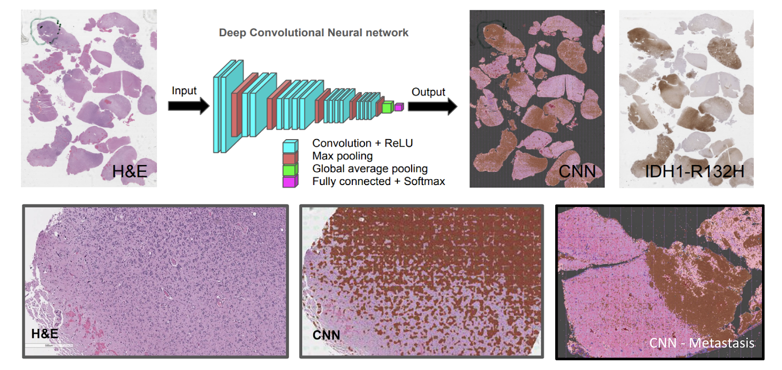 |
| Automated lesion detection and classification in pathology: Upper Panels: A form of artificial intelligence known as deep convolutional neural networks (CNNs) is currently showing impressive results at pattern recognition tasks traditionally carried out by highly skilled humans. In this example, a full digital slide image is analyzed to highlight brain regions infiltrated by this oligodendroglioma. The IDH1-R132H immunostains (right upper panel) highlights where the tumor actually is for comparison. Lower panels: High power view of the same tumor showing it's infiltrative nature. The CNN clearly detects the infiltrative nature of this lesion. The right most panel shows the computer output of another metastatic lesion with a much more circumscribed border. In addition to identifying the lesions, CNNs are beginning to show promise at classifying tumors with different clinical outcomes. |
Monday, June 18, 2018
International Society of Neuropathology set to meet in Tokyo this fall
 |
| Keio Plaza Hotel in Tokyo |
Friday, June 15, 2018
More on the amazing word "physaliferous"
Since my post this past Wednesday about the etymology of the word "physaliferous" which designates the characteristic cells comprising chordomas, the illustrious Dr. Maria Martinez-Lage (neuropathologist at Massachusetts General Hospital), tweeted about another word which derives from the same Greek root. Here is Dr. Martinez-Lage's tweet:
Physaliferous cells resemble the fruit of the physalis plant, an edible berry that is round and surrounded by a delicate lacy husk. It goes by many names: Golden berry, cape gooseberry, edible Chinese lantern. Delicious!

Physaliferous cells resemble the fruit of the physalis plant, an edible berry that is round and surrounded by a delicate lacy husk. It goes by many names: Golden berry, cape gooseberry, edible Chinese lantern. Delicious!

Thursday, June 14, 2018
A Primer on Giant Cell (Temporal) Arteritis
Neuropathologists are often tasked with handling ophthalmic pathology at their institutions. As such, they are assigned all cases submitted by ophthalmologists -- including temporal artery biopsies for determination of the presence of active giant cell (temporal) arteritis. What follows is a quick reference on the important points to remember about giant cell arteritis. (If there are things I am forgetting, please add your comments.):
Refering to the condition as "temporal arteritis" is not entirely accurate as giant cell arteritis is a granulomatous inflammatory disorder that can affect a variety of large and small arteries in the head. In addition to the temporal artery, ophthalmic arteries can be affected (which is the reason ophthalmologists are often the clinicians performing biopsies in suspected cases). Additionally, vertebral arteries and even the aorta (giant cell aortitis) can be involved. Since ophthalmic arteritis can lead to sudden and irreversible blindness, affected patients must be promptly diagnosed and treated. A negative biopsy result does not entirely exclude the diagnosis as the distribution of inflammation is often patchy.
Because pathologic changes tend to be patchy, examination of several cross-sectional levels is required. Involved segments exhibit nodular intimal thickening (and occasional thromboses). Most lesions exhibit granulomatous inflammation within the inner media which disrupts the internal elastic lamina. A minority of cases do not show either granulomas or giant cells, instead exhibiting only a non-specific acute and chronic inflammatory infiltrate. Healing is characterized by intimal thickening, medial thinning, and adventitial fibrosis.
Reference: Kumar V, Abbas AK, and Aster JC (eds.) Robbins Basic Pathology, Chapter 10 "Blood Vessels", 10th Edition (2018) pp. 384-5.
 |
| Arrow points to a giant cell in a temporal artery wall (from Robbins Basic Pathology, 10th edition) |
Because pathologic changes tend to be patchy, examination of several cross-sectional levels is required. Involved segments exhibit nodular intimal thickening (and occasional thromboses). Most lesions exhibit granulomatous inflammation within the inner media which disrupts the internal elastic lamina. A minority of cases do not show either granulomas or giant cells, instead exhibiting only a non-specific acute and chronic inflammatory infiltrate. Healing is characterized by intimal thickening, medial thinning, and adventitial fibrosis.
Reference: Kumar V, Abbas AK, and Aster JC (eds.) Robbins Basic Pathology, Chapter 10 "Blood Vessels", 10th Edition (2018) pp. 384-5.
Wednesday, June 13, 2018
Etymology of the word "physaliferous"
The characteristic cells seen in chordoma, physaliferous cells (which, according to the Oxford English Dictionary, can alternatively be spelled 'physaliphorous') is from the Greek physallis (meaning 'bubble') and phoros (meaning 'bearing').
 |
| The "bubble-bearing" physaliferous cells of a chordoma |
Tuesday, June 12, 2018
A primary central nervous system lymphoma overwhelmed by necrosis and neutrophil infiltration

This PCNSL (later proved to be EBV-driven) biopsied from the right parietal lobe is hardly discernible among the necrotic debris and neutrophilic infiltration
 |
| Tumor cells (outlined) within and surrounding a vessel wall |
Subscribe to:
Posts (Atom)
-
Neuropathology Blog has run its course. It's been a fantastic experience authoring this blog over many years. The blog has been a source...
-
 Shannon Curran, MS with her dissection Shannon Curran, a graduate student in the Modern Human Anatomy Program at the University of Co...