A psychiatrist from the National Bureau of Economic Research in Cambridge, MA, Dr. Karen Norberg spent an entire year knitting detailed replica of the central organ.
I discuss issues pertaining to the practice of neuropathology -- including nervous system tumors, neuroanatomy, neurodegenerative disease, muscle and nerve disorders, ophthalmologic pathology, neuro trivia, neuropathology gossip, job listings and anything else that might be of interest to a blue-collar neuropathologist.
Showing posts with label anatomy. Show all posts
Showing posts with label anatomy. Show all posts
Monday, April 1, 2019
Wednesday, September 12, 2018
Best Post of July 2018: What is the nodulus?
The next in our "Best of the Month" series come from July 16, 2018:
The nodulus is the lobule of the cerebellar vermis that, together with the flocculus of each hemisphere, forms the flocculonodular lobe.
Monday, July 16, 2018
What is the nodulus?
The lobule of the cerebellar vermis that, together with the flocculus of each hemisphere, forms the flocculonodular lobe.
Friday, July 13, 2018
Hyperbrain: a great resource for learning neuroanatomy
 HyperBrain is an online tutorial for human neuroanatomy from the University of Utah. HyperBrain includes thousand of images and hundreds of linked illustrated glossary terms, as well as movies, quizzes and interactive animations.
Monday, May 14, 2018
Understanding Neurophobia Among Medical and Other Health Care Students
 |
| Andre Toulouse, PhD, (University College, Cork, Ireland) lead author on article about neurophobia |
Reference: Javaid MA, Chakraborty S, Cryan JF, Schellekens H, Toulouse A. Understanding neurophobia: reasons behind impaired understanding and learning of neuroanatomy in cross-disciplinary healthcare students. Anat Sci Educ 11:81-93 (2018).
Wednesday, March 14, 2018
On Einstein's Birthday, We Take a Second Look at His Brain
On this date 139 years ago, Albert Einstein was born in Ulm, Germany. We take this occasion to republish a post from November 21, 2012 entitled: Photos reveal unique features of Einstein's cerebral cortex:
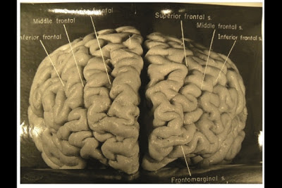
Photographs taken shortly after his death, but never before analysed in detail, have now revealed that Einstein’s brain had several unusual features, providing clues about the neural basis of his extraordinary mental abilities.
Nature.com reports that, while doing Einstein's autopsy, the pathologist Thomas Harvey removed the physicist's brain and preserved it in formalin. He then took dozens of black and white photographs of it before it was cut up into 240 blocks. Now, anthropologist Dean Falk of Florida State University in Tallahassee and her colleagues have obtained 12 of Harvey’s original photographs from the National Museum of Health and Medicine in Silver Spring, Maryland, analysed them and compared the patterns of convoluted ridges and furrows with those of 85 brains described in other studies.Many of the photographs were taken from unusual angles, and show structures that were not visible in photographs that have been analysed previously. The analysis was recently published today in the journal Brain. The most striking observation, says Falk, was “the complexity and pattern of convolutions on certain parts of Einstein's cerebral cortex”, especially in the prefrontal cortex, and also parietal lobes and visual cortex.
The autopsy revealed that Einstein’s brain was smaller than average and subsequent analyses showed all the changes that normally occur with ageing. Nothing more was analysed, however. Harvey stored the brain fragments in a formalin-filled jar in a cider box kept under a beer cooler in his office. Decades later, several researchers asked Harvey for some samples, and noticed some unusual features when analysing them.
A study done in 1985 showed that two parts of his brain contained an unusually large number of non-neuronal cells called glia for every neuron2. And one published more than a decade later showed that the parietal lobe lacks a furrow and a structure called the operculum3. The missing furrow may have enhanced the connections in this region, which is thought to be involved in visuo-spatial functions and mathematical skills.

AFP/Getty Images
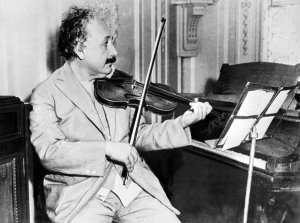
Einstein was a keen violinist, which may account for an overdeveloped section of his brain that deals with the left hand.
The prefrontal cortex is important for the kind of abstract thinking that Einstein would have needed for his famous thought experiments on the nature of space and time, such as imagining riding alongside a beam of light. The unusually complex pattern of convolutions there probably gave the region and unusually large surface area, which may have contributed to his remarkable abilities.
Falk and her colleagues also noticed an unusual feature in the right somatosensory cortex, which receives sensory information from the body. In this part of Einstein’s brain, the region corresponding to the left hand is expanded, and the researchers suggest that this may have contributed to his accomplished violin playing.
According to Sandra Witelson, a behavioural neuroscientist at McMaster University in Hamilton, Canada, who discovered that the parietal operculum is missing from Einstein’s brain, the study’s biggest contribution may be in encouraging further studies. “It makes clear the location and accessibility of photographs and slides of Einstein's brain,” she says. “This may serve as an incentive for other investigations of Einstein's brain, and ultimately of any consequences of its anatomical variations.”
Wednesday, March 7, 2018
The meticulously extracted nervous system of a 19th-century woman on display at Hahnemann Medical College
Last summer I put up a post about a remarkable whole nervous system dissection that was carried out at the University of Colorado School of Medicine. The inimitable Dr. Mark Cohen recently sent me an article about a similar dissection performed at Hahnemann Medical College in Philadelphia by Dr. Rufus B. Weaver. The dissection, which took place in 1888 over the course of five months, was performed on a 35-year-old woman who had given permission for her body to be used for the furtherance of science.
 |
| Dr. Rufus B. Weaver and the nervous system of Harriet Cole |
An excerpt from the article appearing in Atlas Obscura:
According to the History of the Homoeopathic Medical College of Pennsylvania, Dr. Weaver told a fellow doctor about Harriet during a trip to the U.K., after his extraction of the nervous system. He didn’t mention the completion of the dissection. The doctor’s response: “It is impossible, there is no such thing in all this United Kingdom, and if it had been possible it would have been done by some one.” Dr. Weaver replied quietly: “So it has, by some one in the States.”
In an article for Homeopathic World in August 1892, Dr. Alfred Heath was far more generous about Dr. Weaver’s accomplishment. He called it “a marvel of patience and skill in dissection, the likes of which has never been seen before.”
 |
| A dissection similar to that of Dr. Weaver's done at the University of Colorado in 2017 by Shannon Curran |
Thursday, December 14, 2017
Best Post of September 2017 -- Guest Post from Dr. PJ Cimino: Blue discoloration of the gray matter in a patient who received methylene blue for respiratory distress prior to death
The next in our "Best Post of the Month" series is from Monday, September 11, 2017:
Dr. PJ Cimino, whom we profiled when he was a fellow back in November of 2013, is a now faculty member at the University of Washington. I was delighted to receive this email from him today:
"I had an autopsy case with interesting gross pathology findings, which made for some nice clinical images (below). The patient received therapeutic methylene blue in the setting of respiratory distress prior to death. The gross pathology showed striking widespread green-blue gray matter discoloration. I thought these images might be of interest to share with the general neuropatholgy community, and thought your blog might be a good platform to do so, especially since you have posted many good clinical images."
Monday, November 27, 2017
Best Post of June 2017: Remarkable en bloc dissection of human central and peripheral nervous system accomplished at University of Colorado
The next in our "Best of the Month" Series is from June 7, 2017:
 |
| Shannon Curran, MS with her dissection |
Curran, who is known among students and faculty as a preternaturally efficient prosector, completed the dissection in under 100 hours. Further detailed work is planned on the specimen, including dissection of the extraocular muscles away from the eyeballs while maintaining their connection to the brain. Discussion is underway about loaning the specimen to the Denver Museum of Nature and Science for community health education.
 |
| CNS en bloc dissection with extensive portion of PNS |
 |
| Connection to the eyeballs is maintained, with plans to dissect away extraocular muscles |
 |
| Detail showing maintained connection with digital nerves of the left hand |
Monday, September 11, 2017
Guest Post from Dr. PJ Cimino: Blue discoloration of the gray matter in a patient who received methylene blue for respiratory distress prior to death
Dr. PJ Cimino, whom we profiled when he was a fellow back in November of 2013, is a now faculty member at the University of Washington. I was delighted to receive this email from him today:
"I had an autopsy case with interesting gross pathology findings, which made for some nice clinical images (below). The patient received therapeutic methylene blue in the setting of respiratory distress prior to death. The gross pathology showed striking widespread green-blue gray matter discoloration. I thought these images might be of interest to share with the general neuropatholgy community, and thought your blog might be a good platform to do so, especially since you have posted many good clinical images."
"I had an autopsy case with interesting gross pathology findings, which made for some nice clinical images (below). The patient received therapeutic methylene blue in the setting of respiratory distress prior to death. The gross pathology showed striking widespread green-blue gray matter discoloration. I thought these images might be of interest to share with the general neuropatholgy community, and thought your blog might be a good platform to do so, especially since you have posted many good clinical images."
Thursday, June 22, 2017
Best Post of March 2017: Why is the confluence of the cerebral venous sinuses called the "torcula"?
The next in our "Best of the Month" series is from March 3, 2017:
Torcula is derived from a Latin word meaning to “twist” and was also used to refer to a wine press. Within the cranium the venous sinuses come together at the back of the skull in a structure called the confluence of the sinuses. This cavity has four large veins radiating from it, supposedly resembling the spigots that pour dark purple juice out of the four sides of the ancient wine press used to squeeze grapes with a handled screw on the top. The same stem is found in common words such as torture and tortuous.
Monday, June 12, 2017
Additional photograph of remarkable CNS/PNS dissection
Wednesday, June 7, 2017
Remarkable en bloc dissection of human central and peripheral nervous system accomplished at University of Colorado
 |
| Shannon Curran, MS with her dissection |
Curran, who is known among students and faculty as a preternaturally efficient prosector, completed the dissection in under 100 hours. Further detailed work is planned on the specimen, including dissection of the extraocular muscles away from the eyeballs while maintaining their connection to the brain. Discussion is underway about loaning the specimen to the Denver Museum of Nature and Science for community health education.
 |
| CNS en bloc dissection with extensive portion of PNS |
 |
| Connection to the eyeballs is maintained, with plans to dissect away extraocular muscles |
 |
| Detail showing maintained connection with digital nerves of the left hand |
Tuesday, November 24, 2015
The Mercado Brain Cutting Device launched at the University of Colorado
Thanks to a hand-made gift from University of Alabama neuropathology fellow Juan Mercado, MD, our residents on autopsy rotation this month had the opportunity yesterday to inaugurate the use of The Mercado Brain Cutting Device (MBCD). Made from tools easily found at any hardware store, the device allows prosectors to make reliably even 1-cm thick coronal brain slices for optimal demonstration of gross anatomy and pathology. University of Colorado pathology residents Abby Richmond, MD (PGY-III) and Sammie Roberts, MD (PGY-I) used the device to great advantage as demonstrated by the exquisitely presented brain slices laid out for inspection.
Much appreciation to Dr. Mercado for gifting this device, which he describes as a "limited edition (1 or 1)", to our department. We will undoubtedly have more meticulous brain cutting sessions henceforth thanks to Dr. Mercado's efforts.
 |
| Dr. Sammie Roberts with the MBCD |
 |
| Dr. Abby Richmond makes the first cut |
 |
| Sections are even and uniform in thickness |
 |
| The finished product |
 |
| Drs. Moore, Richmond, and Roberts (left to right) examining the coronal sections |
Much appreciation to Dr. Mercado for gifting this device, which he describes as a "limited edition (1 or 1)", to our department. We will undoubtedly have more meticulous brain cutting sessions henceforth thanks to Dr. Mercado's efforts.
Monday, September 28, 2015
The Brain -- as explained by John Cleese
Finally, it has become clear to me now:
Thanks to Dr. Ann Thor for directing me to this concise explanation of the brain and its connections.
Wednesday, September 24, 2014
Best Post of July 2014: 3D print of white matter "captures a sense of delicate complexity that evokes a sense of wonder about the brain"
The next in our "Best of the Month" series is from July 2, 2014:
Creating an accurate 3D model of the brain's white matter for Philadelphia's Franklin Institute was a project no 3D printing company would tackle -- until 3D Systems (Rock Hill, SC) agreed to take it on. Here is an image of the finished project, which took about 210 hours to print out:
Interviewed for the tech website CNET, Franklin Institute chief bioscientist and lead exhibit developer Dr Jayatri Das said that the model "has really become one of the iconic pieces of the exhibit. Its sheer aesthetic beauty takes your breath away and transforms the exhibit space," said . "The fact that it comes from real data adds a level of authenticity to the science that we are presenting. But even if you don't quite understand what it shows, it captures a sense of delicate complexity that evokes a sense of wonder about the brain."
Thanks to the illustrious Dr. Doug Shevlin for informing me of this remarkable feat of engineering which, in his words, sits at "the intersection of neuroscience, computers and 3D printing".
Wednesday, July 2, 2014
3D print of white matter "captures a sense of delicate complexity that evokes a sense of wonder about the brain"
Creating an accurate 3D model of the brain's white matter for Philadelphia's Franklin Institute was a project no 3D printing company would tackle -- until 3D Systems (Rock Hill, SC) agreed to take it on. Here is an image of the finished project, which took about 210 hours to print out:
Interviewed for the tech website CNET, Franklin Institute chief bioscientist and lead exhibit developer Dr Jayatri Das said that the model "has really become one of the iconic pieces of the exhibit. Its sheer aesthetic beauty takes your breath away and transforms the exhibit space," said . "The fact that it comes from real data adds a level of authenticity to the science that we are presenting. But even if you don't quite understand what it shows, it captures a sense of delicate complexity that evokes a sense of wonder about the brain."
Thanks to the illustrious Dr. Doug Shevlin for informing me of this remarkable feat of engineering which, in his words, sits at "the intersection of neuroscience, computers and 3D printing".
Interviewed for the tech website CNET, Franklin Institute chief bioscientist and lead exhibit developer Dr Jayatri Das said that the model "has really become one of the iconic pieces of the exhibit. Its sheer aesthetic beauty takes your breath away and transforms the exhibit space," said . "The fact that it comes from real data adds a level of authenticity to the science that we are presenting. But even if you don't quite understand what it shows, it captures a sense of delicate complexity that evokes a sense of wonder about the brain."
Thanks to the illustrious Dr. Doug Shevlin for informing me of this remarkable feat of engineering which, in his words, sits at "the intersection of neuroscience, computers and 3D printing".
Thursday, June 13, 2013
Best Post of November 2012: Photos reveal unique features of Einstein's cerebral cortex
The next in our "Best of the Month" series is from November 21, 2012:
Photographs taken shortly after his death, but never before analyzed in detail, have now revealed that Einstein’s brain had several unusual features, providing clues about the neural basis of his extraordinary mental abilities.
Nature.com reports that, while doing Einstein's autopsy, the pathologist Thomas Harvey removed the physicist's brain and preserved it in formalin. He then took dozens of black and white photographs of it before it was cut up into 240 blocks. Now, anthropologist Dean Falk of Florida State University in Tallahassee and her colleagues have obtained 12 of Harvey’s original photographs from the National Museum of Health and Medicine in Silver Spring, Maryland, analyzed them, and compared the patterns of convoluted ridges and furrows with those of 85 brains described in other studies. Many of the photographs were taken from unusual angles, and show structures that were not visible in photographs that have been studied previously. The analysis was recently published in the journal Brain. The most striking observation, says Falk, was “the complexity and pattern of convolutions on certain parts of Einstein's cerebral cortex”, especially in the prefrontal cortex, and also parietal lobes and visual cortex.
Wednesday, November 21, 2012
Photos reveal unique features of Einstein's cerebral cortex
Photographs taken shortly after his death, but never before analysed in
detail, have now revealed that Einstein’s brain had several unusual
features, providing clues about the neural basis of his
extraordinary mental abilities.
Nature.com reports that, while doing Einstein's autopsy, the pathologist Thomas Harvey removed the physicist's brain and preserved it in formalin. He then took dozens of black and white photographs of it before it was cut up into 240 blocks. Now, anthropologist Dean Falk of Florida State University in Tallahassee and her colleagues have obtained 12 of Harvey’s original photographs from the National Museum of Health and Medicine in Silver Spring, Maryland, analysed them and compared the patterns of convoluted ridges and furrows with those of 85 brains described in other studies.Many of the photographs were taken from unusual angles, and show structures that were not visible in photographs that have been analysed previously. The analysis was recently published today in the journal Brain. The most striking observation, says Falk, was “the complexity and pattern of convolutions on certain parts of Einstein's cerebral cortex”, especially in the prefrontal cortex, and also parietal lobes and visual cortex.
Nature.com reports that, while doing Einstein's autopsy, the pathologist Thomas Harvey removed the physicist's brain and preserved it in formalin. He then took dozens of black and white photographs of it before it was cut up into 240 blocks. Now, anthropologist Dean Falk of Florida State University in Tallahassee and her colleagues have obtained 12 of Harvey’s original photographs from the National Museum of Health and Medicine in Silver Spring, Maryland, analysed them and compared the patterns of convoluted ridges and furrows with those of 85 brains described in other studies.Many of the photographs were taken from unusual angles, and show structures that were not visible in photographs that have been analysed previously. The analysis was recently published today in the journal Brain. The most striking observation, says Falk, was “the complexity and pattern of convolutions on certain parts of Einstein's cerebral cortex”, especially in the prefrontal cortex, and also parietal lobes and visual cortex.
Thursday, June 14, 2012
Best Post of January 2012: The area postrema is not the only area where the BBB is lacking
The next in our "Best of the Month" series comes from January 9, 2012:
I'll paraphrase a question posed by one of my 2nd-year students at Southern Illinois University School of Medicine:
I'll paraphrase a question posed by one of my 2nd-year students at Southern Illinois University School of Medicine:
I understand that the Area Postrema
was a site where there was increased penetrability of the blood brain barrier.
I am not sure, but thought I had come across additional sites of increased
penetrability last year in my reading. Are there other sites where there is
increased permeability of the BBB?
In pondering an answer to this question, I immediately thought of the
illustrious Dr. John Donahue, consummate neurologist, neuropathologist,
and neuroanatomist. I posed the question to him and got the following
response:
 |
| Dr. John Donahue, Brown University, Providence, RI |
"Not increased permability of the
BBB. NO BBB! Area postrema is one of the circumventricular organs,
areas in the brain that lack a BBB. Being the vomiting center, it is
imperative that it lacks a BBB so that it can sample the systemic circulation.
Being in the medulla, it is the only circumventricular organ that is adjacent to the fourth
ventricle; all of the others are adjacent to the third. It is the only
paired circumventricular organ; all of the others are single and midline.
The other circumventricular organs are subfornical organ, organum vasculosum of the lamina terminalis,
median eminence, posterior pituitary gland, subcommissural organ, and pineal
gland."
There you have it!
Subscribe to:
Posts (Atom)
Neuropathology Blog is Signing Off
Neuropathology Blog has run its course. It's been a fantastic experience authoring this blog over many years. The blog has been a source...
-
Neuropathology Blog has run its course. It's been a fantastic experience authoring this blog over many years. The blog has been a source...
-
A neuropathology colleague in Toronto (Dr. Phedias Diamandis) is developing some amazing AI-based tools for pathology and academia. He hel...


