Showing posts with label muscle. Show all posts

Thursday, March 5, 2020

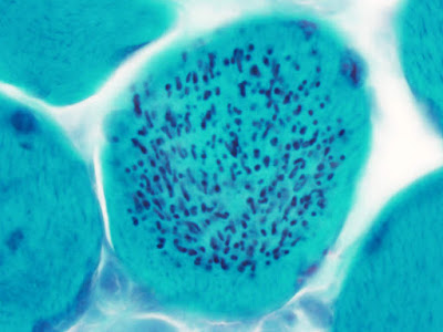
Best Post of October 2019: Cytoplasmic Bodies in a Muscle Biopsy

The next in our "Best of the Month" series comes from October 7, 2019:

The inimitable Dr. Christian Davidson, formerly of Robert Wood Johnson Medical School at Rutgers and now at the University of Utah, sent in pictures of cytoplasmic bodies in a muscle biopsy from a patient with severe myositis. Thank you, Dr. Davidson!

Trichrome

Electron Microscopy

Monday, October 7, 2019

Cytoplasmic bodies in a muscle biopsy

The inimitable Dr. Christian Davidson, formerly of Robert Wood Johnson Medical School at Rutgers and now at the University of Utah, sent in pictures of cytoplasmic bodies in a muscle biopsy from a patient with severe myositis. Thank you, Dr. Davidson!

Trichrome

Electron Microscopy

Friday, March 1, 2019

Donahue does it again: a case of type 2 myotonic dystrophy from Providence, Rhode Island


A case from Dr. John Donahue, inimitable neuropathologist and neuropathology fellowship program director at Brown University:


"This is a fast myosin immunostain on a gluteus maximus biopsy from a 45-year-old woman who went on to have genetically-proven myotonic dystrophy, type 2 (DM2; proximal myotonic myopathy; PROMM).  The type 2 muscle fibers are EXTREMELY atrophic."

Monday, December 10, 2018

A Pattern-Based Approach to the Interpretation of Skeletal Muscle Biopsies

Chunyu (Hunter) Cai, MD, PhD
Chunyu (Hunter) Cai, Douglas Anthony, and Peter Pytel have authored a wonderfully useful review article in Modern Pathology entitled A Pattern-Based Approach to the Interpretation of Skeletal Muscle Biopsies. Dr. Cai wrote the following to me: "In this review, we take a pathology-friendly approach and categorize muscle biopsies into 6  morphologic groups, and discuss common differential diagnoses for each group. I think this review fills a void that there is not a good quick guide for NP fellows, residents and even neuropathologists who don’t sign out muscle cases on a routine base." Thanks, Drs. Cai, Anthony, and Pytel for a review article that will be a fixture on the desks of many neuropathologists in the coming years!

Tuesday, January 16, 2018

Guest Post from Dr. Mike Lawlor: Audentes Announces Positive Interim Data from First Dose Cohort of ASPIRO, a Phase 1/2 Clinical Trial of AT132 in Patients With X-Linked Myotubular Myopathy

Dr. Mike Lawlor
Regular contributor Mike Lawlor, MD, PhD passed this development along from the X-linked myotubular myopathy clinical research front:

Audentes Therapeutics has released an interim data update on the ASPIRO gene therapy clinical trial for X-linked myotubular myopathy.  In the 4th quarter of 2017, three patients were given a single dose of an adeno-associated virus containing the human myotubularin gene.  To quote the press release:

"The early AT132 efficacy data observed in our first dose cohort of patients have exceeded our expectations," stated Dr. Suyash Prasad, Senior Vice President and Chief Medical Officer of Audentes. "At the 12-week timepoint, Patient 1 has improved from a severely compromised baseline to achieve a CHOP-INTEND score and maximal inspiratory pressure that are approaching the ranges normally seen in healthy children.  Importantly, Patient 1 has also attained several age-appropriate developmental milestones within this time period, including head-control, rolling over and sitting unassisted.  While still early in the trial, we view these initial efficacy data as a promising indicator of the potential for AT132 to bring meaningful benefit to patients and families living with this devastating disease."

On the safety side of things, Audentes reported a total of six adverse events (AEs) reported in ASPIRO, two of which were determined to be serious adverse events (SAEs). Per the press release, “Both SAEs occurred in Patient 3, the first of which was a hospitalization one week post-administration due to pneumonia and was deemed not treatment-related. Patient 3 was also hospitalized at week 7 post-administration due to a gastrointestinal infection and elevated troponin levels, the latter of which was deemed probably treatment-related and is responding to treatment with intravenous steroid administration and supportive care.”

On the pathology side of things, it will be a while before biopsies are evaluated or analyzed, as the trial design incorporated extensive strategies to keep the study pathologists blinded to timepoint, treatment group, and even whether a given sample belongs to a study patient.  Hopefully I’ll have the opportunity to share some of the pathology data at the 2019 AANP meeting.


Wednesday, October 11, 2017

Audentes Therapeutics Announces Dosing of First Patient in ASPIRO, a Phase 1/2 Clinical Trial of AT132 for the Treatment of X-Linked Myotubular Myopathy

Following up on the last post, Dr. Mike Lawlor sent me this in an email:

"Audentes Therapeutics officially announced the dosing of the first patient for the X-linked myotubular myopathy treatment trial that we’ve been working on over the past few years.  We’ve been very involved in the translation from dogs to humans, and will be doing the human pathology work for the trial.  Here’s a link to that press release."

Monday, October 2, 2017

Lawlor featured in video about his work in myotubular myopathy

Michael Lawlor, MD, PhD

Medical College of Wisconsin is highlighting the work of our colleague Dr. Michael Lawlor in the area of gene therapy for myotubular myopathy. Check out this wonderful 4-minute video!

Monday, January 23, 2017

FDA approves first drug for spinal muscular atrophy

The U.S. Food and Drug Administration recently approved Spinraza (nusinersen), the first drug approved to treat children and adults with spinal muscular atrophy (SMA). The FDA granted this application fast track designation and priority review. The drug also received orphan drug designation, which provides incentives to assist and encourage the development of drugs for rare diseases.

Thanks to Dr. Nick Willard for alerting me to this development.

Wednesday, April 16, 2014

Dr. Mike Lawlor is recognized for making critical strides in treatment of myotubular myopathy

X-linked myotubular myopathy (XLMTM) is a severe and fatal congenital myopathy for which there is currently no treatment.  As part of an international group of scientists studying treatments for this disease, Dr. Mike Lawlor (the neuropathologist serving Children's Hospital of Wisconsin) was recently featured on Milwaukee's WTMJ 10 o'clock news for his part in the work.  The piece is quite touching. Mike conveys a sense of urgency in finding a cure for this disease because he has gotten to
know several children stricken with XLMTM, and even has pictures of many of the little tikes up on his office wall.  Mike has presented his work on XLMTM at the past several AANP meetings, including trials using myostatin inhibition, targeted enzyme replacement therapy, and gene therapy on animal models of this disease.  The work in this story relates to the gene therapy trial that was recently published in Science Translational Medicine (depicted on the left), for which Mike was the study pathologist. A photomicrograph from the article was even featured on the front page of that prestigious journal.  He is continuing to work on these treatment options in preparation for translation to human clinical trials that will occur over the next few years. Congratulations to Dr. Mike Lawlor for splendidly representing neuropathologists in both the lay media and academic press.

Friday, June 28, 2013

Dr. Mike Lawlor's Proposed Muscle Biopsy Checklist


Comments are encouraged!!!!


Clinical History
1.  Gender of patient:    __male ___female
2.  Age at presentation:  _ _ years _ _ months
3.  Age at biopsy: _ _ years _ _ months
4.  Symptoms at presentation (check all that apply):
 Weakness
 Hypotonia
 Muscle pain
 Cardiac disease
 Central nervous system disease
 Respiratory difficulties
 Contractures
 Failure to thrive
 Others (see item 8)
5.  Elevated creatine kinase:  Yes  No  Unknown _______ Patient Value _________(Normal Range)
6.  Familial Inheritance:____None      ___Autosomal Recessive     ____Autosomal Dominant     ____X-linked
7.  EMG Findings: ____Not known        ____Myopathic              ____Neuropathic
8.  Other symptoms, signs, and lab data: ____________________________________________________________________________________________________________________________________________________________________________________________________________________________________________________________________________________

Muscle Biopsy Tissue Information

1.            Name of Muscle:___________________________________________________________________

2.            Size of tissue collected*:  ______  X  ______   X  ______ cm

3.            Date of tissue collection*:        __ __ / __ __ / __ __ __ __
                                                m  m     d   d     y   y   y   y

4.            Biopsy method:  Open         Needle


5.            Freezing or Fixation Used*?      Frozen:                   Amount: _____ mg     Not known
 Formalin-fixed:         Amount: _____ mg     Not known
 Paraffin-embedded: Amount: _____ mg     Not known      
 Epon-embedded:      Amount: _____ mg     Not known

Histological Findings in Muscle Biopsy or Autopsy specimens

1.       Which standard histochemical stains were used*? (choose all that apply)
 H and E                   Gomori trichrome      NADH         COX            SDH
 COX/SDH                PAS                        Oil Red O     ATPase 4.3  ATPase 4.6
 ATPase 9.4              Other, specify: __________________________________________________________________________________________________________________________________________________________________________________

2.       Which of the following diagnostic abnormalities were noted on histochemical stains (choose all that apply)*?
Fatty replacement                ___absent         ___mild             ___moderate                 ___severe
Endomysial fibrosis              ___absent         ___mild             ___moderate                 ___severe
Myofiber degeneration                      ___absent         ___mild             ___moderate                 ___severe
Necrosis                             ___absent         ___mild             ___moderate                 ___severe
Myophagocytosis                 ___absent         ___present in ____ fibers
Myofiber regeneration (Basophilic fibers)        ____absent       ___present in _____ fibers
Abnormalities of fiber type          ____absent       _____present
Specify*:      Type 1 predominance                       ______ % Type 1 fibers
 Type 2 predominance                 ______% Type 2 fibers
 Fiber type grouping (of both fiber types)
Hypertrophic fibers               ____absent       _____present in _____ fibers
Atrophy/Hypotrophy                   ____absent       _____present
Specify:       All fibers within the specimen          
 Subsets of fibers, leading to excessive variation in fiber size
Specify (choose all that apply):    Single fibers                         Groups of fibers      
                                     Type 1 fibers only     Type 2 fibers only
Perifascicular distribution
             Atrophic/hypotrophic fiber shape
 Angulated     Round    
Myopathy-associated pathological structures, specify:     
Central nuclei                _____absent      _____present
Specify estimated % of fibers (include eccentric nuclei): _____
            Internal nuclei                _____absent      _____present
Specify estimated % of fibers (if not quantified above): _____
Inclusion bodies             ____absent       ____present in _____ fibers
Rimmed vacuoles                      ____absent       ____present in _____ fibers
            Nemaline rods               ____absent       ____present
Specify:             Restricted to one fiber type, specify which: _____     
                                                 Nuclear rods present
Ragged red fibers                      ____absent       ____present in _____fibers
COX- negative fibers                  Estimated number ______
Strongly SDH-reactive blood vessels (SSV’s)                  _____absent      _____present
Central cores                 ____absent       ____present in _____ fibers
            Minicores                      ____absent       ____present in ____ fibers
            Core-like lesions            ____absent       ____present in ____ fibers
            Targetoid fibers              ____absent       ____present in ____ % of fibers
            Marked hypotrophy of type 1 fibers                      ____absent       ____present
Inflammation                 ___absent         ___mild             ___moderate                 ___severe
Specify:
 Distribution
 Perivascular
                               Evidence of vascular damage            Thrombi identified in blood vessels
                         Focal
 Diffuse
 Endomysial
 Perimysial
 Involving fascia
             Associated with myofiber damage
              Associated with non-necrotic myofiber
              Granulomas
                        Necrotizing   Non-necrotizing   Giant cells present    Foreign material present
             Inflammatory cells identified
                        Specify (choose all that apply):
                         Lymphocytes
                         Neutrophils
                         Macrophages
                         Eosinophils (as a prominent component)
                         Microorganisms identified, specify which: _________________________________________
Abnormal storage material
Specify:
                        Excessive glycogen                    ____absent      ____mild           ____severe
                        Excessive intracellular lipid         ____absent       ____mild           ____severe
Additional observations
____________________________________________________________________________________________________________________________________________________________________________________________________________________________________________________________________________________

3.       Which immunohistochemical stains were used? (choose all that apply)
 Myosin immunohistochemistry (for fast and slow fibers)         
 Dystrophin panel
            Specify:
                        Dystrophin (DYS1)         ____absent       ____reduced      ____normal
                        Dystrophin (DYS2)         ____absent       ____reduced      ____normal
                        Dystrophin (DYS3)         ____absent       ____reduced      ____normal
                        Dystrophin (BMD Hotspot)         ____absent       ____reduced      ____normal
                        Spectrin                                    ____absent       ____reduced      ____normal
                        Utrophin                                    ____absent       ____normal       ____increased
                       
 Other stains for limb-girdle or congenital muscular dystrophy
            Specify:           
                        Laminin a2/Merosin                   ____absent       ____reduced      ____normal
                        Alpha dystroglycan (VIA)            ____absent       ____reduced      ____normal
                        Alpha dystroglycan (IIH)             ____absent       ____reduced      ____normal
                        Beta dystroglycan                      ____absent       ____reduced      ____normal
                        Alpha sarcoglycan                     ____absent       ____reduced      ____normal
                        Beta sarcoglycan                       ____absent       ____reduced      ____normal                  
Delta sarcoglycan                      ____absent       ____reduced      ____normal
                        Gamma sarcoglycan                  ____absent       ____reduced      ____normal
Dysferlin                                   ____absent       ____reduced      ____normal                  
Emerin                                      ____absent       ____reduced      ____normal
                        Collagen VI                               ____absent       ____reduced      ____normal
                        Caveolin 3                                 ____absent       ____reduced      ____normal
                        Desmin                                     ____absent       ____reduced      ____normal
                        Integrin a7                                ____absent       ____reduced      ____normal
                        nNOS                                       ____absent       ____reduced      ____normal
                       
 Inflammatory myopathy panel
                        CD4                                         ____absent       ____present in ___ % of lymphocytes
                        CD8                                         ____absent       ____present in ___ % of lymphocytes
                        CD20                                        ____absent       ____present in ___ % of lymphocytes
                        CD45                                        ____absent       ____present in ____% of mononuclear cells        
CD68                                        ____absent       ____present in ____% of mononuclear cells        
C5b-9                                       ____absent       ____present on endomysial capillary walls
Major Histocompatability Complex          ____absent     ____sarcolemmal       ____diffuse

4.        Additional immunohistochemical/immunofluorescence assays performed: __________________________________________________________________________________


5.       Other abnormalities noted on immunohistochemistry:  __________________________________


Epon-Embedded Tissue/Electron Microscopy (Muscle Biopsy/Autopsy Specimens)

1.       Abnormalities seen on:             Light microscopy (Toluidine blue staining)       Electron microscopy
 Both – Light microscopy and Electron microscopy

2.       Abnormalities noted in:            Contractile apparatus
                                                 Sarcotubular organization
                                                 Mitochondria, specify (choose all that apply):
 Abnormal shape       
 Abnormal numbers
                                                                         Abnormal location   
                                                                         Abnormal architecture

3.       Describe any pathological inclusions noted:     N/A _________________________________________________________________________________________

_________________________________________________________________________________________

4.       Describe any abnormal storage material identified:       N/A _________________________________________________________________________________________

_________________________________________________________________________________________


Interpretation:


Comment:


Neuropathology Blog is Signing Off

Neuropathology Blog has run its course. It's been a fantastic experience authoring this blog over many years. The blog has been a source...