Monday, December 31, 2012

Best Post of July 2012: "Chasing The Dragon": A Cause of Toxic Spongiform Leukoencephalopathy

The next in our "Best of the Month" series comes from July 10, 2012:


My favorite case from the 2012 AANP Diagnostic Slide Session in Chicago last month featured an autopsy slide from the brain of a 25-year-old man with a history of polysubstance abuse found unresponsive at a New Year's Eve party. Toxicology screening was positive for methadone, lorazepam, and cocaine. The patient died after three weeks in the intensive care unit. Attendees were provided glass slides in advance of the session demonstrating the following findings:
Low Power: Marked white matter pallor

High Power: White matter virtually replaced by lipidized macrophages
Presenter Joshua Menke, MD of The Mayo Clinic (Rochester, MN) revealed that white matter damage was diffuse, including infratentorial structures. The subcortical U-fibers were relatively spared and myelin was disproportionately affected as compared to axons, as demonstrated in these photomicrographs from Dr. Menke's presentation:
LFB stain on left and neurofilament stain on right
Discussants included Drs. Tessa Hedley-Whyte, Craig Horbinski, Mark Cohen, and others. Before the diagnosis was revealed, Dr. Horbinski stated that he thought Delayed Hypoxic Leukoencephalopathy might be the best fit for this case. Dr. Cohen pointed out that Delayed Hypoxic Leukoencephalopathy tends spare the infratentorial regions, while the white matter damage is more diffusely distributed in toxic leukoencephalopathy. This case was ultimately revealed to be that of Toxic Spongiform Leukoencephalopathy.

Toxic leukoencephalopathy can be caused by a range of insults including radiation, chemotherapy, and drugs of abuse. Among the drugs of abuse that have been shown to cause toxic leukoencephalopathy are toluene, ethanol, cocaine, 3,4-methylenedioxy-methamphetamine, intravenous heroin, inhaled heroin pyrolysate, and psilocybin (Ref: Filley & Kleinschmidt-DeMasters NEJM 2001). Toxic spongiform leukoencephalopathy is a clinicopathologic entity first associated with the inhalation of pyrolysate heroin vapors, a practice which had its origins in Hong Kong in the 1950's and which came to be known colloquially as "chasing the dragon". Users typically "chase the dragon" by placing heroin on a creased piece of tin foil over a flame. As the drug sublimates, the user inhales the fumes. The pathophysiology of this form of leukoencephalopathy has not been clearly elucidated, but is thought to be related to a direct toxic effect of heroin on oligodendrocytes.

Friday, December 21, 2012

Best Post of June 2012: Neuropathologists party hard at AANP Annual Reception

The next in our "Best of the Month" series comes from June 22, 2012 and recalls fond memories of an excellent annual meeting of the American Association of Neuropathologists this past summer in Chicago:

L to R: Drs. Heather Sumner and T. David Bourne
Drs. Mark Cohen, T. David Bourne, and Mahtab Tehrani
L to R: Drs. Edward Stopa, William Taylor, and Qian Wu


Friday, December 14, 2012

What is this vagus stuff?

Henry A. Lester, PhD
Actually, it's Vagusstoff (literally translated from German as "Vagus Substance") refers to the substance released by stimulation of the vagus nerve which causes a reduction in the heart rate. Discovered in 1921 by physiologist Otto Loewi, vagusstoff was the first confirmation of chemical synaptic transmission and the first neurotransmitter ever discovered. It was later confirmed to be acetylcholine, which was first identified by Sir Henry Hallett Dale in 1914. Because of his pioneering experiments, in 1936 Loewi was awarded the Nobel Prize in Physiology or Medicine, which he shared with Dale. Thanks to Dr. Henry Lester of Cal Tech for informing me of this interesting bit of neuro-history through his free online course entitled "Drugs and The Brain". The information provided here is taken from the Wikipedia article on the topic.

Tuesday, December 4, 2012

"the NFL is a breeding ground for mental illness" -- Former Denver Bronco Nate Jackson

Today, a friend forwarded me this opinion piece about the murder-suicide of NFL linebacker Jovan Belcher. I responded that I blogged about such cases before, and I didn't intend to write yet another blog post about chronic traumatic encephalopathy. I felt as though lethal violence among football players is happening so often now, it's not even worth yet another post. But, the fact that the violence has reached this level of banality is in itself worthy of comment. Don't worry, though, the next time an NFL player commits homicide or suicide, I promise not to write about it.

Wednesday, November 21, 2012

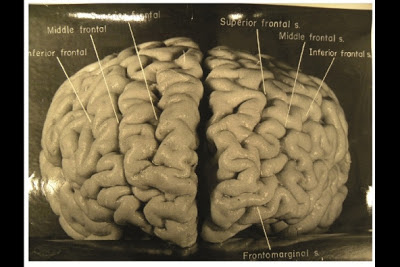
Photos reveal unique features of Einstein's cerebral cortex

Photographs taken shortly after his death, but never before analysed in detail, have now revealed that Einstein’s brain had several unusual features, providing clues about the neural basis of his extraordinary mental abilities.


Nature.com reports that, while doing Einstein's autopsy, the pathologist Thomas Harvey removed the physicist's brain and preserved it in formalin. He then took dozens of black and white photographs of it before it was cut up into 240 blocks. Now, anthropologist Dean Falk of Florida State University in Tallahassee and her colleagues have obtained 12 of Harvey’s original photographs from the National Museum of Health and Medicine in Silver Spring, Maryland, analysed them and compared the patterns of convoluted ridges and furrows with those of 85 brains described in other studies.Many of the photographs were taken from unusual angles, and show structures that were not visible in photographs that have been analysed previously. The analysis was recently published today in the journal Brain. The most striking observation, says Falk, was “the complexity and pattern of convolutions on certain parts of Einstein's cerebral cortex”, especially in the prefrontal cortex, and also parietal lobes and visual cortex.

Thursday, November 15, 2012

CAP seeking input on revised brain/spinal cord tumor protocol


College of American Pathologists LogoPrior to the release of the revised Brain/Spinal Cord Protocol, the College of American Pathologists (CAP) will host a public comment period. You can review the revisions and submit feedback online. The public comment period will run through November 30, 2012. Please note that comments are not instantly available for review and will be posted on a weekly basis.

Tuesday, November 13, 2012

Review Article: "Thinking and Talking About Life Expectancy in Incurable Cancer"

As a follow-up to my previous post, here's a perspective from a June 2011 article in Seminars in Oncology:

"Most patients with incurable cancer want information about the impact cancer will have on their future, and many want specific estimates of the most likely, best case, and worst case scenarios for survival. With improved understanding of life expectancy, patients are better equipped to make appropriate treatment decisions and plans for the future. Although physicians acknowledge that patients with incurable cancer want prognostic information and benefit from this, most struggle to provide it and experience difficulty in making reliable estimates, communicating them, and tailoring the information to the individual patient."

Thursday, November 8, 2012

Why genetically profile a glioma?

A couple of weeks ago I met with representatives of Castle Biosciences regarding their proprietary gene expression profile assay for glioblastomas, called DecisionDx-GBM, as well as their multi-methylation test for grade II and III gliomas, called DecisionDX-G-CIMP. During the meeting, I brought up a crucial issue raised by a Neuropathology Blog reader. I'll quote part of the reader's comment: "I doubt anyone wants to give a patient a life expectancy prediction based on the results of this test, since individuals may fall at any point on a survival curve. So I would not be eager to recommend this test until there are alternative treatments for those in poor prognosis groups." The representative's response to this concern was that prognostication is an important part of the decision-making process that a neuro-oncologist, in collaboration with the patient, takes into account in recommending a course of treatment. For example, when should Avastin be introduced into the regimen? If a patient has a tumor with a genetic signature that has a longer median survival, perhaps the neuro-oncologist would be more likely to hold back on the introduction of Avastin, keeping it in his armamentarium for later in the disease course. I would add that if I personally had the misfortune of being diagnosed with a glioblastoma, I would like to have as much prognostic information as possible just to help me adjust psychologically to dealing with the the disease. I realize that one individual can fall at any point on a survival curve. But put yourself in the patient's shoes for a moment. Wouldn't you want this test done, as well as IDH1 mutation, MGMT methylation status, 1p/19q testing, and whatever else that might be available to get as much information as possible about this disease has invaded you? Since, as Castle Biosciences states, insurance will cover this test, I see both practical and intangible benefits to doing it. I would be curious to hear what other neuropathologists have to say about this issue in general and about the Castle Biosciences tests in particular. I should note that the Castle tests were developed by the widely-respected neuropathologists Dr. Kenneth Aldape at MD Anderson. I am seriously entertaining the idea of routinely using the Castle Biosciences tests at my institution and would be interested in the advice of the neuropathology community as a whole regarding this issue. Please post to comments.

Tuesday, November 6, 2012

Best Post of May 2012: New Study Looks at Head Impacts in Youth Football

The next in our "Best of the Month" series is from May 14, 2012:

We neuropathologists, and society as a whole, have spent a lot of time over the last couple of years rethinking the long-term effects of repeated concussive and subconcussive blows to the heads of professional and college football players. But what about the 3.5 million kids in American who play below the high school level? Dr. Peter Cummings today sent me a link to a report regarding groundbreaking research being done at Virginia Tech in which impact-measurement instruments were placed on 7 and 8-year-old football players. Data was collected on more than 750 hits to the head over the course of the season.


Lead researcher, Stefan Duma, a professor of Biomedical Engineering, reports that some head impacts in youth football are equal in force to some of the bigger hits he sees at the college level.  The average kid received 107 head impacts during the course of the season. This is fewer than seen in high school (which averages about 500 per season) and college (which amounts to about 1000 per season.). But what about the magnitude of these hits? The median impact was 15 g's. In Duma's study, there were 38 impacts that were 40 g or greater (almost all of which occurred during practice). Six impacts were over 80 g's, which starts to get into the range of risk for concussion. “Nobody expected to see hits of this magnitude,” says Duma, who speculates that once players start seeing hits of 30 g's or above, there is a risk for cumulative injury.

Here's the video report from reporter Stone Philips. The interviews with the parents of these youth players perplexed me. All of the parents featured found the results concerning, but none expressed any thoughts about having their child switch to another sport.

Thursday, November 1, 2012

Robert Scully Has Died


A message just received from neuropathologist David Louis, Pathologist-in-Chief at Massachusetts General Hospital:

Dr. Robert E. Scully
Dear colleagues,
It is with deep sadness that I let you know that our dear friend, mentor and colleague, Dr. Robert E. Scully, passed away late yesterday afternoon [October 30] at the age of 91.

Dr. Scully was a giant in the field of pathology who devoted almost all of his career to the Mass. General Hospital. He was on the active staff for 55 years and stepped down from active practice only in the past decade. After graduating from Harvard Medical School in 1944, he trained at the Peter Bent Brigham Hospital and then joined our staff in the early 1950s. Dr. Scully rapidly became known as a peerless diagnostic pathologist. Although he soon became recognized around the world for special expertise in gynecologic and testicular pathology, his opinion was sought in nearly all areas of pathology by colleague after colleague for many decades here at MGH and elsewhere. He contributed over 480 papers to the literature and described many hitherto unrecognized entities, largely in gynecologic pathology but also to a significant degree in testicular pathology. His book on the ovary was considered the standard reference work on the subject and the current classification of gynecologic tumors derives largely from his work. Dr. Scully was also the Editor of the Case Records of the Massachusetts General Hospital in New England Journal of Medicine for a remarkable 27 years. He was an Emeritus Professor of Pathology at Harvard Medical School.

Dr. Scully was also a gentleman and a gentle man. He was quiet but firm, honest, careful, considerate and happy to give credit to others. As such, he was a beloved figure in the field of pathology worldwide.


Thursday, October 25, 2012

Netter Pituitary Hormone Illustration

If you're delivering a lecture on the pituitary gland, you might find this illustration useful to include in your PowerPoint presentation:

Monday, October 22, 2012

IDH1 Mutations in Oligodendrogliomas: PNA-clamping PCR is best analytic method

This is just out from a Korean group headed by Dr. Se-Hoon Kim publishing online in Brain Pathology:

Although direct sequencing of mutations in isocitrate dehydrogenase 1 (IDH1) has been considered to be the gold standard method to detect this mutation, the sensitivity of this technique has been questioned especially because specimens from glial tumors may contain large numbers of non-tumor cells. The group screened 141 cases of oligodendroglial tumors for IDH1 mutations using peptide nucleic acid (PNA)-mediated clamping PCR and compared the results with the results of direct sequencing, pyrosequencing, and immunohistochemistry (IHC). Nested PCR was only performed in cases having mutant IDH1 only discovered by clamping PCR. Using dilution experiments mixing IDH1 wild-type and mutant DNA samples, clamping PCR detected mutations in samples with a 1% tumor DNA composition. Using PNA clamping PCR, the group detected 138 of 141 (97.9%) cases with mutant IDH1 in the series, which is significantly higher (P = 0.016; PNA clamping vs. direct sequencing) than those of direct sequencing (74.5%), pyrosequencing (75.2%), and IHC (75.9%). From these results, it appears that almost all oligodendroglial tumors have IDH1 mutations, suggesting that IDH1 mutation is an early and common event especially in the development of oligodendroglial tumors.

Thursday, October 18, 2012

Dramatic meningioma manifestations

Orbitocranial extension of meningioma
Massive cranial extension
Nasal extension of meningioma
Massive frontal lobe meningioma

Monday, October 1, 2012

A 32-year-old HIV+ male presenting with mental status changes

 Soon after admission, the patient lapsed into a coma and died.

Minimal edema (brain weight: 1500 grams)

Some hypoxic-ischemic damage as evidenced by Purkinje red neuron change
"Soap bubble" changes around some vessels (sample from basal ganglia)
Giant cells among mixed inflammatory infiltrate in the meninges
Inflammation appears to extend minimally into brain parenchyma
Organisms consistent with crytopcoccus neoformans amid inflammation
Mucicarmine-positive fungi capsules highlighted in red, with some capsules partially deficient
Diagnosis: Cryptococcal meningoencephalitis with early hypoxic-ischemic encephalopathy.

Wednesday, September 26, 2012

Microcystic meningioma in the frontal lobe of a 57-year-old female

When confronted by giant, pleomorphic nuclei among rather non-meningothelial cells, as depicted below, the neuropathologist given a specimen purported to be a benign meningioma might hesitate before agreeing with the neuroradiologist's and neurosurgeon's presumptive diagnosis.
But in this case, a rather small meningotheliomatous component was present to provide reassurance:
A glimpse of a shattered psammoma body also helped to confirm that this was indeed a grade I meningioma, with a predominantly microcystic component.
The microcystic variant is among the most common of the "nonclassical" meningiomas.

Friday, September 21, 2012

Best Post of the April 2012: Photomicrographs of spinocerebellar ataxia type 6 case

The next in our "Best of the Month" series is from April 26, 2012. It's a follow-up from a prior post in which I showed the gross finding from the same case of spinocerebellar ataxia type 6:

A month ago, I posted gross photographs from an autopsy case of mine of a patient with spinocerebellar ataxia type 6. I am now following up with histologic images for this case. The most striking feature was the marked loss of Purkinje cells, which makes sense because SCA6 involves mutation of a calcium channel that is highly expressed on Purkinje neurons:
Many remaining Purkinje neurons demonstrated degenerative changes, including mis-shapened nuclei
Attenuated cerebellar granule cell layer

More Purkinje cells in bad shape

Cerebellar dentate neurons also dengenerated, but not as extensively as Purkinje cells


Mild involvement of inferior olives. Note the mis-shapen and darkened nuclei of neurons in this location.

Pons minimally effected, with only occasional degenerating neurons as seen in the center of this photomicrograph next to a normal neuron

Neuropathology Blog is Signing Off

Neuropathology Blog has run its course. It's been a fantastic experience authoring this blog over many years. The blog has been a source...