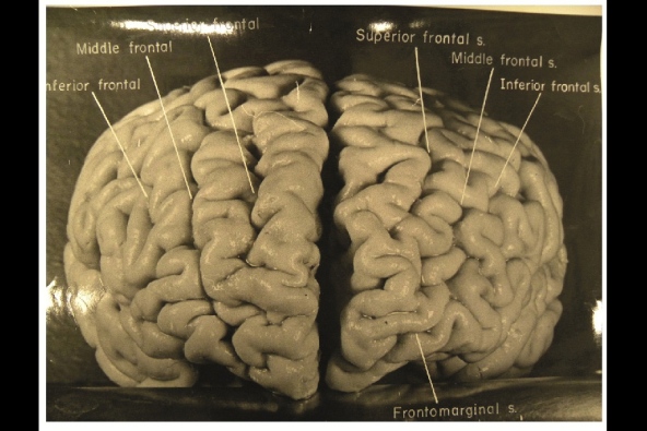
Nature.com reports that, while doing Einstein's autopsy, the pathologist Thomas Harvey removed the physicist's brain and preserved it in formalin. He then took dozens of black and white photographs of it before it was cut up into 240 blocks. Now, anthropologist Dean Falk of Florida State University in Tallahassee and her colleagues have obtained 12 of Harvey’s original photographs from the National Museum of Health and Medicine in Silver Spring, Maryland, analysed them and compared the patterns of convoluted ridges and furrows with those of 85 brains described in other studies.Many of the photographs were taken from unusual angles, and show structures that were not visible in photographs that have been analysed previously. The analysis was recently published today in the journal Brain. The most striking observation, says Falk, was “the complexity and pattern of convolutions on certain parts of Einstein's cerebral cortex”, especially in the prefrontal cortex, and also parietal lobes and visual cortex.

No comments:
Post a Comment