
This translucent mass was identified in the third ventricle:

Histological examination of the cyst contents revealed:

Although no ciliated epithelium was identified within the specimen, there were columnar cells with a pseudostratified configuration:
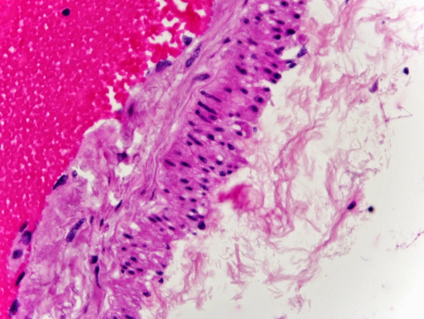
However, the majority of the cyst lining was flattened, simple epithelium:

Diagnosis? (Please post to comments)

9 comments:
Diagnosis is colloid cyst of the third ventricle. Normally one would see ciliated columnar cells lining the cyst, but they appear to be attenuated and flattened by the internal pressure of the cyst itself.
Brian. there a group on Facebook you might be interested in.
America's Colloid Cyst Surviors.
May I know th size of the cyst?
To "Houston Genaux", thank you, I will check that FaceBook page out.
To "Premyscake", the cyst was 1.1 centimeter in greatest dimension.
thanks Brian, your going to have to ask to join....no problem there which you will be our first to involved with us.
Which I will explain Facebook graph to you and how to use it as a tool for research.
You can ask the group multiple choice question by 'ask questions' option also.
thanks Brian, your going to have to ask to join....no problem there which you will be our first to involved with us.
Which I will explain Facebook graph to you and how to use it as a tool for research.
You can ask the group multiple choice question by 'ask questions' option also.
There is a larger group on Facebook called Colloid Cyst Survivors which is worldwide. My daughter was diagnosed with a colloid cyst at a young age and is being monitored with annual MRIs to track it's growth. We are in Canada and I think neuros take a wait and see approach to them here rather than operate right away. So far it has not grown at all and is 7mm. She is almost 17 yrs old. She is not symptomatic. It is my opinion that these are more common than we previously thought. Now that more people are having CT scans done for concussions etc they are showing up more often and at a younger age.
How long do they usually wait to remove the cyst, and is it possible to just let it be
To Mary Petrucha,
Surgical management is handled on a case-by-case basis. There is no way to tell how a particular case should be handled. It is possible to just let such a cyst alone if it is not causing any problems for the patient.
Post a Comment