Wednesday, December 18, 2013

Best Post of September 2013

The next in our "Best of the Month" series is from September 16, 2013

Two UC Davis Neurosurgeons Resign After Intentionally Infecting Intracranial Glioblastomas with Bowel Bacteria

The Sacramento Bee recently reported that two UC Davis neurosurgeons who intentionally infected three glioblastoma patients with bowel bacteria have resigned their posts after the university found they had "deliberately circumvented" internal policies, "defied directives" from top leaders and sidestepped federal regulations, according to newly released university documents.

Neurosurgeon J. Paul Muizelaar, MD

Dr. J. Paul Muizelaar, 66, the former head of the neurosurgery department, and his colleague, Dr. Rudolph J. Schrot, violated the university's faculty code of conduct. All three patients consented to the procedures in 2010 and 2011. Two of the patients died within weeks of their surgeries, while the other survived more than a year after being infected.

Read more here: http://www.sacbee.com/2013/08/25/5678851/uc-davis-surgeons-resign-after.html#storylink=cpy

Muizelaar and Schrot called their novel approach "probiotic intracranial therapy," or the introduction of live bowel bacteria, Enterobacter aerogenes, directly into their patients' brains or bone flaps. The doctors theorized that an infection might stimulate the patients' immune systems and prolong their lives. The first patient lived about 5 1/2 weeks. The second survived another year, an outcome that buoyed the doctors and seemed to bolster their theory, they said. The institutional trouble began in March 2011, when a newly diagnosed third patient developed sepsis, became unresponsive and died two weeks after being deliberately infected. The university's first internal investigation soon followed.

Muizelaar and Schrot did not complete a study on rats before they began treating the three human patients, despite an FDA directive in 2008 that "animal studies will be necessary prior to entering into the clinic with your proposed therapy," according to an email to Schrot from an FDA official. When asked by a compliance investigator why animal trials were not done first, Schrot allegedly responded that such testing would take "10 years … his entire career," one internal review states. The investigator found Schrot's "eagerness to proceed" to be concerning and his actions "reckless."


Muizelaar, one of the highest paid employees in the University of California system, stepped down effective June 27. He earned more than $907,000 last year, the 33rd-highest gross pay in the UC system.  Schrot, 45, an associate professor who made about $512,000 last year, will leave his post at the end of August. Both doctors have sharply criticized the findings, characterizing the internal investigations as biased and incomplete. The surgeons maintain they were acting in the best interests of their desperately ill patients, whose prognoses for survival were poor. Both doctors opted not to appeal the university's findings because they determined it would be fruitless."I lost confidence, if you will, in the ability of the university administration to fairly handle it," Schrot said Friday in the office of his Sacramento attorney.


Read more here: http://www.sacbee.com/2013/08/25/5678851/uc-davis-surgeons-resign-after.html#storylink=cpy
One colleague, Dr. David Asmuth, an infectious disease specialist who co-chaired an oversight committee for university research, called the surgeons' procedure "the worst case of human subjects research he had ever seen."

Read more here: http://www.sacbee.com/2013/08/25/5678851/uc-davis-surgeons-resign-after.html#storylink=cpy

"I was simply thinking that I could help patients," Muizelaar said. "My whole medical practice is guided by actually only one principle, namely: What would I do for my mother, my son, myself?"

Tuesday, December 10, 2013

The Solution to the Football Concussion Dilemma: Hold All Games at High Altitude

A new study shows that high school athletes playing at higher altitudes suffer fewer concussions than those closer to sea-level, a phenomenon attributed to physiological changes in the brain causing it to fit more tightly in the skull.

"This is the first time any research has linked altitude to sports-related concussion," said Dawn Comstock, PhD, associate professor of epidemiology at the Colorado School of Public Health and co-author of the study. "It appears that when you are at altitude there may be a little less free space in the skull so the brain can't move around as much."

The study, first-authored by David Smith, MD, of Cincinnati Children's Hospital Medical Center, was published recently in the Orthopaedic Journal of Sports Medicine.

The researchers analyzed concussion statistics from athletes playing multiple sports in 497 high schools from across the U.S. with altitudes ranging from 7 feet to 6,903 feet with 600 feet being the median. They also examined football separately since it has the highest concussion rate of high school sports. The numbers came from the National High School Sports-Related Injury Surveillance System directed by Comstock.
The results showed a 31 percent decrease in concussion rates among all high school sports played at altitudes of 600 feet and above. Concussion rates for high school football players at these altitudes decreased by 30 percent.

"Vasogenic edema in the brain leads to increased extravascular water," the study says. "These two adaptations would also lead to a tighter packaging of the brain with increased blood cell content surrounding the brain."
 
"If this study is correct, we should look to replicate our findings in the National Football League," Comstock said. "For example, if the Broncos play the Chargers in San Diego or the Dolphins in Miami they should experience more concussions than when they play here in Denver."

The incidence of concussion among high school athletes has grown tremendously. The Centers for Disease Control and Prevention estimate the annual incidence of sports-related traumatic brain injury in the U.S. at 1.6 million to 3.8 million with many more going undiagnosed. In a recent 10-year period there has been a 100 percent increase among 8 to 13-year-olds and a 200 percent increase among 14 to 19-year-olds in sports-related emergency room visits for concussion.

Interestingly, scientists found that putting mild pressure on a rat's jugular vein increased pressure on the brain and reduced injury from concussion by 83 percent.

Thanks to the esteemed Dr. Doug Shevlin, pictured below, for alerting me to this remarkable finding.

Douglas Shevlin, MD


Tuesday, December 3, 2013

What happened to neuropathology in 1946?

The esteemed Dr. Jim Mandell and his son were recently playing with the amazing Google Books Ngram Viewer. Google Ngram searches a huge corpus of books for the mention of a particular search term. It then graphs the frequency with which that term appears over time. On a whim, the Mandell's entered the search term "neuropathologist". Here's the resulting graph:

I couldn't fit the y-axis label in the picture, but it ranges from 0% up to 0.00000300%. Dr. Mandell challenges his neuropathology colleagues to explain the sharp spike in the usage of "neuropathologist" around 1946-47. Please enter your speculations in the comment section. Dr. Mandell also points out the lamentable fact that it "appears we are past our peak".

Friday, November 22, 2013

Best Post of August 2013: Profile of Wash U NP Fellow Dr. PJ Cimino

The next in our "Best of the Month" series is from August 1, 2013, when I profiled Dr. PJ Cimino, a prominent first-year neuropathology fellow at Washington University in St. Louis. After a short biographical sketch, Dr. Cimino answers a few of my questions:


Dr. PJ Cimino

P.J. Cimino grew up in Seattle, WA, where he did both his undergraduate studies (double major in neurobiology and biochemistry) and Medical Scientist Training Program (combined MD/PhD program) at the University of Washington. He earned his PhD in Neurobiology and Behavior while working in the laboratory of Tom Montine MD, PhD in the Department of Pathology. His graduate work mainly focused on the biology of microglia related to prostaglandin signaling and neurodegenerative disease. He graduated in 2011 and moved his family from Seattle to the Midwest to train in the combined Anatomic Pathology (AP)/Neuropathology (NP) program at Washington University in St. Louis, MO. He has completed hist first two years of AP training and is now in the NP fellowship portion. During his time in St. Louis he has also developed a budding interest in molecular neuro-oncology. In addition to gaining experience in diagnostic neuropathology, he plans to return to the laboratory bench in order to continue on his track to become an independent physician-scientist in investigative neuropathology. P.J. is married to Heather, and they have three children: Sadie, Dominic, and Francis.
1.       What does the PJ stand for?
Patrick Joseph.
2.       Why did you decide to become a neuropathologist?
The short answer is that neuropathology just fits with my personality and interests. As an undergraduate I was fascinated by neurologic disease and worked in a laboratory that studied the genetics of inherited neurological disorders. After making the decision to become a physician-scientist, I knew that I was drawn to the field of neurology. As many people of my ilk do, I spent time exploring neurosurgery, neurology, psychiatry, neuropathology, etc. I was fortunate enough as a graduate student to join the lab of a great neuropathologist, who showed me what neuropathology was like. After exploring all of those 'neuro' options, neuropathology was the right fit for me. I think it will afford me the opportunity to study the mechanism of neurologic disease while having a hand in patient care.
3.       Name a couple of important professional mentors. Why were they important to you?
Beginning in chronological order, I first have to recognize my undergraduate research mentor at the University of Washington, Wendy Raskind MD, PhD. She was the first person to inspire me to become a physician-scientist. I saw how she managed patients clinically as well as ran a basic science research lab, and used these two endeavors to complement and enhance one another. The next important mentor has to be Tom Montine MD,PhD, as mentioned above. He initially got me interested in neuropathology, and essentially helped me to solidify my career goals. He has helped me tremendously over the past several years and still provides much needed sound guidance. In the past couple of years I have gained some newer and emerging professional mentors at Washington University in St. Louis, including BobSchmidt MD, PhD and David Gutmann MD, PhD. I do want to acknowledge that this list is not comprehensive and I am a product of several other great professional mentors that I have met along the way.
4.       What advice would you give to a pathology resident interested in doing a neuropathology fellowship?
Do it! I have met academic neuropathologists and private practice pathologists who have completed neuropathology training. So there appears to be many career choices for those with neuropathology fellowship training. I think that the vast majority of neuropathologists (again in my experience) need to have a skill in addition to neuropathology. So in addition to NP training, you should consider either doing another pathology fellowship (clinical track) or a post-doctoral fellowship/mentored research (research track).  You just have to take into consideration your personal and professional goals and plan your training appropriately. For full disclosure to residents, I cannot provide comprehensive advice about neuropathology and what lies beyond fellowship training, because I am still in training myself. Hopefully, I will get a 'real job' someday as an academic neuropathologist and I can add to this advice in the future.
5.       What city would you like a future American Association of Neuropathologists meeting to be held and why?
I think that any city in the Pacific Northwest or Mountain region would be a good place to hold a national meeting that takes place in the end of June. These include: Denver, Seattle, Portland, Salt Lake, etc. I think that the end of June is a good time to head North and West to cool down for a few days while looking at posters, going to talks, and attending the diagnostic slide session. The organizers must have anticipated my response to this question, as they have preemptively scheduled the 2014 meeting to take place in Portland, OR.

Thursday, November 7, 2013

Best Post of July 2013: New Muscle Pathology Text edited by Goebel, Sewry, and Weller is available

This post, which origninally appeared on July 11, 2013, is being re-posted as part of the "Best of the Month" series for those who would consider purchasing it now that it is available on the market. Amazon is selling the paper version for $229.48, and a Kindle edition for $124.99.

The second edition of Muscle Disease: Pathology and Genetics will be released in August 2013. The publisher states that the book "clarifies the pathology and genetics of muscle disease for pathologists, clinicians, geneticists and researchers to aid in the diagnosis and management of patients. Organized around the 'motor unit' concept, this book presents the latest understanding of muscle disease, and how this can help identify new treatments."

Friday, November 1, 2013

Nanotechnology joins with cancer genomics in silencing glioblastoma oncogene

Gold nanoparticles (yellow) with small interfering RNAs (green) knock down an oncogene in glioblastoma.
In a study of mice released this week in Science Nanomedicine, researchers were able to reduce glioblastoma size three- to four-fold by switching off the oncogene Bcl2Like12 by means of nanotechnology-assisted delivery of small interfering RNAs. Normal (linear) nucleic acids cannot get into cells, but these spherical nucleic acids can. Small interfering RNA (siRNA) surrounds a gold nanoparticle like a shell; the nucleic acids are highly oriented, densely packed and form a tiny sphere. (The gold nanoparticle core is only 13 nanometers in diameter.) The RNA’s sequence is programmed to silence the disease-causing gene.

“This is a beautiful marriage of a new technology with the genes of a terrible disease,” said Chad A. Mirkin, a nanomedicine expert and a senior co-author of the study. “Using highly adaptable spherical nucleic acids, we specifically targeted a gene associated with GBM and turned it off in vivo. This proof-of-concept further establishes a broad platform for treating a wide range of diseases, from lung and colon cancers to rheumatoid arthritis and psoriasis.”

Dr. Alexander H. Stegh discovered the Bcl2Like12 oncogene in 2007.  "The beauty of the gene we silenced in this study is that it plays many different roles in therapy resistance,." says Stegh. "Taking the gene out of the picture should allow conventional therapies to be more effective.”

Again, thanks to loyal reader and friend, Dr. Doug Shevlin (pictured below on far left with alt country singer Eef Barzelay on far right), for alerting me to this new development in the field of nanotechnogenomics.

L to R: Dr. Doug Shevlin, John Jordan, and alt country legend Eef Barzelay

Wednesday, October 30, 2013

Guest Post from Dr. Keith Kaplan: Why Pathology?

The following post appeared yesterday in Dr. Keith Kaplan's Digital Pathology Blog. I thought the message was important enough to re-post it here:

“What animates a great pathologist? Is it the desire to cure disease, to save life? Surely not, save perhaps as an afterthought. He is too intelligent, deep in his soul, to see anything praiseworthy in such a desire. He knows from life-long observation that his discoveries will do quite as much harm as good, that a thousand scoundrels will profit to every honest man, that the folks who most deserve to be saved will probably be the last to be saved. … What actually moves him is his unquenchable curiosity—his boundless, almost pathological thirst to penetrate the unknown, to uncover the secret, to find out what has not been found out before.”
— H. L. Mencken

Why did I choose pathology?  Why does anyone choose pathology as a medical specialty?
Keith Kaplan, MD
Some of my thoughts on this were previously shared in “What a Beautiful Sea of Granulomas” this summer.

For me it was about the sense of discovery, the sense of making the unknown, known and helping people through the right diagnosis for the right treatment. As H.L. Mencken puts it, “to find out what has not been found out before”.

Why did you choose pathology?  What are your frustrations?  Would you choose medicine as a career now and would you still choose pathology? – Contact me or comment with your thoughts.

In medical school, the chair of pathology spoke to us the first day of second year in our first pathology didactic lecture and began his lecture with a transparency on an overhead projector (remember that?) by saying that “The hospital is the church, the pathologist is the priest”.  The next transparency went on to mention that the liver is the most important organ in the body saying there are brainless, gutless and heartless people but there are no liverless people, therefore the liver was the most important organ.  His interests and grant dollars focused on liver disease.

What about now?  In 2013, days from more announcements about more reimbursement cuts for pathology tests and services in 2014?  What motivates medical students to choose pathology?  Or will they avoid entering the field, instead drawn to specialties that society appears to value more if you measure value in CPT codes and reimbursements?

A well-known pathologist recently gave me the three reasons he went into and continues to practice pathology:
1.  90% of the work Pathologists do is interesting, only 10% is boring (Clinicians have the opposite %s)
2.  Can work in any area of medicine and with a broad focus across many organs, vs a clinician specialist in a single organ
3.  I don’t have to worry about non-compliant patients, i.e. patients not taking their medicine, etc.

And when asked about current frustrations in hospital-based Pathology:
1.  Lack of resources – bean counters slow progress for critical new initiatives
2.  We don’t get the attention/public attention for our work or value. Pathology is performed behind the scenes
3.  There’s WAY too much work

Another academic pathologist offered:
1. Pathology would still be my first choice in medicine due to the unique blend of imaging, daily challenging diagnostic dilemmas and access to and experimentation with cutting edge technologies almost on a continual basis.
2. One of the hidden exceptional values of Pathology as a specialty is the time it affords you to lead a balanced and fulfilling life as the demands are not nearly as pressing as those in other disciplines particularly surgery and medicine, yet you have potentially much more of a chance to impact patient lives if you do your job effectively and speak often to the clinicians and patients you serve (and not only with your reports).
3. Finally, pathology done effectively lets you directly be involved in many of the critical decisions that affect a patient’s life through continually educating those around you and at the same time being a vanguard for proper care delivery.  Done well you have the privilege of being a “doctor’s doctor”.  All other jobs and careers I have seen (both inside and outside of medicine), nothing compares to Pathology in terms of daily impact.  Want to have an impact?  Become a pathologist…the opportunities to lead are myriad.

That being said, this is what is holding Pathology back today:
1. Pathology needs to integrate instead of silo the many exciting moving parts under one roof.  Anatomic and Clinical Pathology are just too fragmented and need to work together more effectively and focus on patient centric reporting instead of a diarrhea of separate disconnected values.  Put the thinking back in our practice and focus on training pathologists as knowledge engineers, and de-emphasizing the toll booth mentality (for one piece of data you make sure you charge and get reimbursed for one RVU and you produce one report).
2. The Pathology societies, AACC, AABB, AANP, AMP, API, ASCP, CAP, USCAP and others, must work more effectively together to create “one international voice” for our specialty instead of focusing on their niche messages.  Today more than ever Pathology needs a clear message not white noise and cacophony of niche interests.  Every pathologist should attend the largest medical meeting in the world, the Radiology Society of North America (average attendance 87,000 with over 50% international attendees).  The brute force Radiology has accomplished with RSNA needs to happen in Pathology.  We pathologists can actually learn a lot from one another.
3. We need to attract the best and the brightest to our amazing discipline.  This should start with “pipeline” programs bringing high school students to our laboratories and then inviting them back summer after summer during their college and medical school years to do research and experience the pure joy of microscopy, advanced blood based diagnostics and pathology applied research that abound in each and every one of our pathology labs.  By opening the eyes of the best and the brightest to our discipline, perhaps we can start getting excited ourselves again about the magic that lies within our practice.  We need leadership and visionaries at the helm to ensure a bright future for Pathology and we need to start now.

My responses to the positives and negatives are below:
1.  I love the process of continual discovery that Pathology provides. Last week at a small hospital I saw for the first time in the operating room a patient with 4 ovaries.  Few specialties offer seeing something new daily.
2.  There is a very wide and endless variety in the work.
3.  I’m fascinated by histology.  The art of taking human tissue and processing it and sectioning it to make a slide and a diagnosis.
Current frustrations:
1.  Lack of resources
2.  Low professional stature
3.  Society doesn’t value Pathologists or the work we do

Why did you choose pathology?  What are your frustrations?  Would you choose medicine as a career now and would you still choose pathology? – Contact me or comment with your thoughts.
 

Would like to compile results and summarize in a future post what you like about your practice and your frustrations.

Tuesday, October 22, 2013

New study suggests "glymphatic system" flushes brain of toxins during sleep

Douglas Shevlin, MD
A new study published in the journal Science on Thursday and reported in the Washington Post suggests that a so-called "glymphatic system" seems capable of flushing toxins (including perhaps beta-amyloid) from brain -- particularly during sleep.“Sleep puts the brain in another state where we clean out all the byproducts of activity during the daytime,” said study author and University of Rochester neurosurgeon Maiken Nedergaard. (Thanks to avid NP Blog reader and friend, Dr. Doug Shevlin, for alerting me to this significant new finding.)

Wednesday, October 16, 2013

Duke Docs say: Don't ignore microvasculature in evaluating muscle biopsies

Dr. Anne Buckley
Drs. Anne Buckley and Edward Bossen of Duke University have a nice review article in the current issue of Journal of Neuropathology and Experimental Neurology entitled Skeletal Muscle Microvasculature in the Diagnosis of Neuromuscular Disease. Buckley and Bossen bring attention to the often overlooked blood vessels in skeletal muscle biopsies. They state that "there are many vascular features in skeletal muscle biopsies that, when interpreted in the context of other histologic patterns and clinical history, provide useful information that allows muscle pathologists to narrow their differential diagnoses and provide more accurate guidance to treating physicians." In addition to discussing normal and abnormal muscle vascular histology, the authors discuss the vascular effects of factors common to many patients undergoing muscle biopsy, such as diabetes mellitus, hypertension, and aging.
Capillary loss (B) and gain (D) in different myopathic processes (DM=dermatomyositis)

Thursday, October 10, 2013

Best Post of June, 2013: Mutations in COQ2 in Familial and Sporadic Multiple System Atrophy

The next in our "Best of the Month" series is from June 15, 2013:

Researchers from the Multiple System Atrophy (MSA) Research Collaborative in Japan just published online in the New England Journal of Medicine findings providing evidence that functionally impaired variants of the COQ2 gene (involved in the biosynthetic pathway for coenzyme Q10) are associated with an increased risk of developing MSA. This group previously identified multiplex families with MSA, indicating a genetic component in a disease that had previously been considered a non-genetic disorder.

Thursday, October 3, 2013

Best Post of May 2013: Angulated intramitochondrial inclusions in the left leg of a man with a left-sided limp

Here's the next in our Best of the Month Series. No definitive diagnosis has been suggested since the original post on May 9, 2014:

A 59-year-old male with a one-year history of limping as well as pain, weakness, and paresthesia of the left lower extremity who underwent lumbar microdiscectomy with cord decompression. Although the patient's pain subsided post-operatively, his other symptoms persisted. Since his CPK levels were chronically elevated (around the 500's), biopsies were later performed on the quadriceps bilaterally. Light microscopic examination was underwhelming except for some denervation effect on the left. However, subsarcolemmal clumping was evident with NADH histochemisty on both the right and left. This clumping prompted me to perform an ultrastructural examination, which revealed the following angulated intramitochondrial inclusions IN THE WEAK LEG ONLY:


These inclusions were present in about 5% of mitochondria.  I reported out my findings in a descriptive manner, not certain of their significance. If anyone has seen such intramitochondrial inclusions in a specimen, please comment. As of now, the patient has no definitive diagnosis.

Wednesday, September 25, 2013

Thirteen patients in New England put at risk for iatrogenic CJD

CNN reports that a patient who had undergone neurosurgery in New Hampshire later developed autopsy-confirmed sporadic Creutzfeldt-Jakob disease (sCJD). Before the patient's disease was discovered, the same nonsurgical equipment used on the CJD patient was used on thirteen subsequent patients, putting those patients at risk for prion infection. The Centers for Disease Control has said that no cases of the disease linked to the use of contaminated medical equipment have been reported in the United States since 1976.

Monday, September 16, 2013

Two UC Davis Neurosurgeons Resign After Intentionally Infecting Intracranial Glioblastomas with Bowel Bacteria

The Sacramento Bee recently reported that two UC Davis neurosurgeons who intentionally infected three glioblastoma patients with bowel bacteria have resigned their posts after the university found they had "deliberately circumvented" internal policies, "defied directives" from top leaders and sidestepped federal regulations, according to newly released university documents.

Neurosurgeon J. Paul Muizelaar, MD

Dr. J. Paul Muizelaar, 66, the former head of the neurosurgery department, and his colleague, Dr. Rudolph J. Schrot, violated the university's faculty code of conduct. All three patients consented to the procedures in 2010 and 2011. Two of the patients died within weeks of their surgeries, while the other survived more than a year after being infected.

Read more here: http://www.sacbee.com/2013/08/25/5678851/uc-davis-surgeons-resign-after.html#storylink=cpy

Muizelaar and Schrot called their novel approach "probiotic intracranial therapy," or the introduction of live bowel bacteria, Enterobacter aerogenes, directly into their patients' brains or bone flaps. The doctors theorized that an infection might stimulate the patients' immune systems and prolong their lives. The first patient lived about 5 1/2 weeks. The second survived another year, an outcome that buoyed the doctors and seemed to bolster their theory, they said. The institutional trouble began in March 2011, when a newly diagnosed third patient developed sepsis, became unresponsive and died two weeks after being deliberately infected. The university's first internal investigation soon followed.

Muizelaar and Schrot did not complete a study on rats before they began treating the three human patients, despite an FDA directive in 2008 that "animal studies will be necessary prior to entering into the clinic with your proposed therapy," according to an email to Schrot from an FDA official. When asked by a compliance investigator why animal trials were not done first, Schrot allegedly responded that such testing would take "10 years … his entire career," one internal review states. The investigator found Schrot's "eagerness to proceed" to be concerning and his actions "reckless."


Muizelaar, one of the highest paid employees in the University of California system, stepped down effective June 27. He earned more than $907,000 last year, the 33rd-highest gross pay in the UC system.  Schrot, 45, an associate professor who made about $512,000 last year, will leave his post at the end of August. Both doctors have sharply criticized the findings, characterizing the internal investigations as biased and incomplete. The surgeons maintain they were acting in the best interests of their desperately ill patients, whose prognoses for survival were poor. Both doctors opted not to appeal the university's findings because they determined it would be fruitless."I lost confidence, if you will, in the ability of the university administration to fairly handle it," Schrot said Friday in the office of his Sacramento attorney.


Read more here: http://www.sacbee.com/2013/08/25/5678851/uc-davis-surgeons-resign-after.html#storylink=cpy
One colleague, Dr. David Asmuth, an infectious disease specialist who co-chaired an oversight committee for university research, called the surgeons' procedure "the worst case of human subjects research he had ever seen."

Read more here: http://www.sacbee.com/2013/08/25/5678851/uc-davis-surgeons-resign-after.html#storylink=cpy

"I was simply thinking that I could help patients," Muizelaar said. "My whole medical practice is guided by actually only one principle, namely: What would I do for my mother, my son, myself?"

Read more here: http://www.sacbee.com/2013/08/25/5678851/uc-davis-surgeons-resign-after.html#storylink=cpy

Read more here: http://www.sacbee.com/2013/08/25/5678851/uc-davis-surgeons-resign-after.html#storylink=cpy


Read more here: http://www.sacbee.com/2013/08/25/5678851/uc-davis-surgeons-resign-after.html#storylink=cpy


Read more here: http://www.sacbee.com/2013/08/25/5678851/uc-davis-surgeons-resign-after.html#storylink=cpy

Read more here: http://www.sacbee.com/2013/08/25/5678851/uc-davis-surgeons-resign-after.html#storylink=cpy

Wednesday, September 11, 2013

Best Post of April 2013: A Rich Focus

The next in our "Best of the Month" series is a post from April 16, 2013.

The grey arrow is pointing to a Rich focus that has hemorrhaged into the subarachoid space
A Rich focus is a tuberculoma in the cerebral cortex. Rich foci become particularly significant when they rupture into the subarachnoid space and cause tuberculous meningitis. This entity is named for Johns Hopkins pathologist Dr. Arnold Rice Rich (1893-1968), who first described it.

Wednesday, September 4, 2013

Dr. Marta Couce joins neuropathology staff at Case Western

Dr. Marta E. Couce

Marta E. Couce, MD, PhD, will be joining the staff of University Hospitals of Cleveland, Case Western Reserve University  next week. She will teaming up to handle surgical neuropathology with the illustrious Dr. Mark Cohen, who has been on staff at Case since 1993.

Dr. Couce attended Medical School in Santiago de Compostela, Spain. She pursued her graduate school training at the same University in the Department of Pathology.  After research stints at East Carolina University and the Mayo Clinic, Dr. Couce did her training in Anatomic Pathology at Yale New Haven Hospital, followed by two fellowships - in Surgical Pathology and Neuropathology - at Mayo Clinic in Rochester, Minnesota. She worked as a staff Neuropathologist at UPMC, in Pittsburgh, leaving in 2004 to join the Department of  Anatomic Pathology at Son Espases University Hospital in Mallorca, Spain, where she became Chair of Anatomic Pathology, a position that she held until her departure in August, 2013.

Her main research interests are metabolic neuropathology and brain tumors. She has held grants, both in the US and  Spain, to study the effects of diabetes and obesity in the brain and for the last 8 years has been involved in the study of oxidative stress and territorial differences in gliomas.

She is be joined in the bustling metropolis of Cleveland by her husband, an endocrinologist at Cleveland Clinic, and her two teenaged children.

Monday, August 26, 2013

Best Post of March 2013: An oddball sellar region mass

The next in our "Best of the Month" series is from Friday, March 15, 2013:


In the last post, it was noted that Dr. Peter Burger presented a series of sellar region "oddball lesions" at the recent USCAP meeting. The esteemed Dr. Mark Cohen was good enough to provide photographs (above) of one particular lesion that was discussed at the meeting: an osteolipoma of the tuber cinereum. Thank you, Dr. Cohen!

Friday, August 16, 2013

What's on your tumor biomarker wish list?

Photo courtesy of Shellie Sherrod
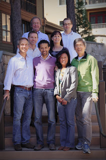
The College of American Pathologists Neuropathology Committee (pictured, minus the illustrious Dr. Aaron Wagner) poses the following question to the neuropathology community worldwide:

"If you were constructing a panel of eight biomarkers there were well-established prognostic/predictive markers for CNS neoplasms, what markers would be included? Note: this is not for diagnosis, but for predicting behavior and should include adult and pediatric brain tumors and can include immunohistochemical and molecular tests."

Thanks in advance for providing your opinion in the comments section.

Tuesday, August 13, 2013

Best Post of February 2013: Review by Dr. Mark Cohen of "Neuropathology: A Volume in the Hight Yield Pathology Series", edited by Yachnis and Rivera-Zengotita

The next in our "Best of the Month" series appeared on February 26, 2013:


Mark L. Cohen, MD
I am honored to present a guest post by the inimitable Dr. Mark Cohen of the illustrious Case Western Reserve University. Dr. Cohen not only reviews a great new neuropathology textbook, but illustrates yet again why he is widely known as the Maxwell Smart of Neuropathology.


(Not really) Full disclosure
Self-annihilating conflicts of interest, as follows:
  1. Long-standing professional relationship with unbridled admiration for lead editor Tony Yachnis, both as a person and as a pathologist (he's not the Moderator of the world-famous Diagnostic Slides Session of the American Association of Neuropathologists, as well as their President-Elect, for nothing).
  2. I have contributed to several publications that compete within the same niche, and from which I have amassed a small fortune (recently enabling me to purchase new windshield wipers for my ‘99 Corolla).
Several weeks ago, I received the following teaching evaluation from an anonymous medical student: "One of the worst lecturers that I had the misfortune of experiencing in medical school. Powerpoints were poorly constructed in terms of high yield content...”. So, when our host Dr. Moore blogged about the impending publication of Neuropathology: A Volume in the High Yield Pathology Series, I sprinted to my laptop to pre-order a copy.
Anthony T. Yachnis, MD, MS
The book arrived safely last night (albeit buried in several inches of snow), and I was not disappointed. Best described as Text-Atlas, it weighs in at 351 pages, which is roughly half that of both Perry & Brat’s Practical Surgical Neuropathology and Prayson’s Neuropathology, second edition. In addition, it represents something of a revolutionary approach in that it was primarily written by trainees in pathology, neuropathology, dermatopathology, hematopathology and neurosurgery. This team-based approach (a testament to Captain Tony’s coaching skills) has produced a neuropathology text “of the people, by the people, and for the people”, as well as resulting in a textbook which is remarkably up to date (including such topics as Natalizumab-related PML). In addition to an introduction to basic neuropathological reactions, subjects covered include developmental disorders (both malformative and acquired), cerebrovascular disorders, trauma, brain tumors, infectious diseases, neurodegenerative disorders, demyelinating diseases, toxic/metabolic disorders, and neuromuscular disorders. For a book of this size, the scope of the topics covered is remarkably comprehensive, and they are extensively, if not exhaustively, illustrated. The pictures, especially the gross photographs, are of uniformly high quality (which appears to be maintained in the on-line version, unlike another high-profile textbook I reviewed a few years ago). In addition to the beautifully photographed gross specimens, pictures include histochemistry, immunohistochemistry, and neuroimaging, as appropriate. The vast majority of the photographs are reproduced at 3.25 x 2.5 inches, which is more than adequate to appreciate the features described in the figure legends. With rare exceptions, arrows are not included within the photomicrographs (which I feel is appropriate, but has gotten me in trouble with certain journals, publishers, and students). As is the practice within this series of textbooks, as well as in many other currently published pathology books, the text is entirely bulleted (a practice which I personally dislike, but which is executed quite well here). Another feature that the reader should be aware of is that (no doubt as part of the philosophy of “high-yield pathology”) there are no references.
Marie Rivera-Zengotita, MD
As an academic neuropathologist, do I have criticisms? (Do bears sh...?). Besides minor quibbles with organization (I probably would have included Natilizumab-associated PML within the Infections section rather than as part of multiple sclerosis) and such Talmudic issues as whether brain invasive meningiomas are truly atypical or not really atypical, but still WHO Grade II, I think that the improvements that I would suggest are largely outside the purview of the authors and editors. Specifically, the titling typography and design layout is nothing short of hideous, and, although the pictures are of adequate size, the presence of many nearly blank pages certainly makes me wish that those spaces had been filled by larger format photographs. In addition, a few of the topics are presented sans photographs, which detracts somewhat from the flow of the textbook (although the authors had the good sense of not trying to illustrate mixed oligoastrocytomas). All in all, however, I think that the book succeeds in its aspirations of presenting "high yield neuropathology".
The editors hope that the volume will be useful for trainees in pathology, neurology, neurosurgery, neuroradiology, neuro-oncology and related fields; as well as being a practical reference for practicing pathologists -- including neuropathologists requiring quick access to the field. As I was reading through the text, I kept this in mind, and triaged these groups as follows:
  1. Neurology and neurosurgery residents should run, not walk, to order a copy, as Board examinations are near at hand, and this book provides a perfect study guide for preparation. Neuropathology fellows should also obtain a copy for studying, although at this moment they have time to walk to their laptops or bookstore.
  2. Neuroradiologists and anatomic pathologists should obtain a copy to keep as a quick reference
  3. Neuro-oncologists will probably gravitate towards more specialized textbooks (such as the recent addition to the Diagnostic Pathology series edited by PCB et al.)
  4. As far as practicing neuropathologists go, I think we all need a copy as we generally find ourselves in one of two situations:
    1. A private practice or group setting without instantaneous access to literature databases. For this group, I think the book fulfills its goal as a quick access to diseases with which we may have become unacquainted during our time in practice.
    2. A large academic medical center with broadband access to literature databases. In this position, our quick access will generally be on-line. On the other hand, we should all have a copy to lend to clinical rotators as an expression of our great goodwill. (Another minor weakness of the book is that there is no RFID device by which we can geolocate our books once they have left our offices. On the other hand, the included on-line access assures that even as our copies disappear, we will still have access from our computers).
Unfortunately, while I feel this book will definitely help to guide me in the revision of my medical student lectures, it is by itself not appropriate for medical student study. To paraphrase Jack Nicholson as Col Nathan R. Jessup, USMC in A Few Good Men: You want high yield? You can’t handle high-yield!
Luckily, for those who can handle the truth, Neuropathology: A Volume in the High Yield Pathology Series will provide a valuable and lasting resource.

Neuropathology Blog is Signing Off

Neuropathology Blog has run its course. It's been a fantastic experience authoring this blog over many years. The blog has been a source...