 |
| Will Smith portraying Bennet Omalu, MD in the movie "Concussion" |
Wednesday, September 2, 2015
Neuropathology Goes Hollywood: "Concussion" Opens in Theaters December 2015
Dr. Bennet Omalu, the first neuropathologist to draw a connections between playing football and the development of Chronic Traumatic Encephalopathy, is featured in a movie starring Will Smith slated to open in December. Check out the trailer!
Not surprisingly, according to a New York Times report, the National Football League intervened in the making of the film so that the final cut would not vilify the nation's most-watched game.


Sunday, August 23, 2015
Best Post of January 2015: Dr. Pierluigi Gambetti steps down as director of national prion surveillance center
The next in our "Best of the Month" series comes from January 5, 2015:
 |
| Pierluigi Gambetti, MD |
"I will be resigning as Director of the National Prion Disease Pathology Surveillance Center effective January 1, 2015. I will continue to be associated with the Center in an advisory position and as consultant for special cases. Dr. Jiri Safar, Associate Professor of Pathology and Neurology at Case Western Reserve University, will be the new director."
Dr. Gambetti goes on to state that the NPDPSC "will remain unchanged", continuing to coordinate autopsies on suspected prion disease cases. Dr. Mark Cohen will continue to have primary responsibility for the histologic and immunohistochemical assessment of cases.
Saturday, August 15, 2015
College of American Pathologists Neuropathology Committee Meets this Weekend
I am delighted to be in Chicago this weekend meeting with my wonderful colleagues on the College of American Pathologists Neuropathology (CAP-NP) Committee. We are making plans for a SAM-eligible educational product that will update you on the 2015 iteration of the World Health Organization Classification of Tumors of the Central Nervous System. The new WHO book is set to be published in October of this year; and we on the committee are making plans to create a CD to be issued a year from now designed to keep you in the loop regarding the latest in CNS tumor classification. This weekend's meeting also marks the end of Dr. Dan Brat's tenure as CAP-NP chairman. Dr. Brat will be replaced by the illustrious Dr. Eyas Hattab at the helm of the CAP-NP Committee. After a long day at work today on the CAP-NP educational product, committee members retired to Smith and Wollensky Steakhouse for some well-deserved nourishment before returning to finish up our work tomorrow. In addition to Drs. Brat, Hattab, and myself, current committee members include Drs. Bill Hickey, Joe Ma, Roger McLendon, Matthew Schneiderjan, Aaron Wagner, Cynthia Welsh, and junior member Matthew Cykowski.
 |
| Rania Hattab (wife of Dr. Eyas Hattab) and Dr. Joe Ma enjoy a morsel of chocolate cake at tonight's CAP-NP dinner |
 |
| Outgoing CAP-NP committee members Dr. Dan Brat and Dr. Cynthia Welsh will be sorely missed |
Wednesday, August 5, 2015
The Tumor Biomarker Series: INI1
This is the last in my tumor biomarker series -- at least until future significant biomarkers are established. I conclude this series with a short description of INI1, a marker for atypical teratoid/rhabdoid tumor (AT/RT). A clinically aggressive embryonal tumor of infancy, AT/RT is characterized by mutations in SMARCB1/INI1 (HSNF5). Immunohistochemical evaluation of AT/RT for the INI1 protein using the BAF47 antibody shows a loss of labelling in tumor cell nuclei, with retention of staining in internal positive control cells such as endothelial cells. Since AT/RT has morphologic overlap with medulloblastoma, CNS PNET, choroid plexus carcinoma, GBM, and other malignant tumors of childhood, INI1 immunohistochemistry is extremely useful in arriving at a diagnosis of AT/RT. A diagnosis of AT/RT carries implications for genetic counseling as this tumor -- in about a one-third of cases -- is a component of the rhabdoid tumor predisposition syndrome (RTPS) wherein there is a germline mutation of SMARCB1/INI1. Because of the risk associated with RTPS, the germline status of SMARCB/INI1 is typically assessed for each new case of AT/RT.
Wednesday, July 29, 2015
Best Post of December 2014: The Einstein/Rorke-Adams Connection
The next in our "Best of the Month" series comes from Tuesday, December 16, 2014:
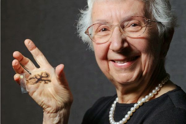 |
| Dr. Lucy Rorke-Adams |
Friday, July 17, 2015
The Tumor Biomarker Series: Medulloblastoma Markers
Four subgroups of medulloblastoma have been defined based on genetic alterations:
Wingless (WNT) - WNT medulloblastomas display monosomy 6 and most show nuclear accumulation of the WNT pathway protein beta-catenin, which serves as a useful immunohistochemical screen for this group. Medulloblasomas with more than 50% nuclear staining for beta-catenin have been shown to have WNT pathway activation, whereas those with only focal nuclear staining do not. Overall survival for WNT medulloblastomas are dramatically longer than those of other subtypes, and clinical practices surrounding the treatment of this subtype reflects this better prognosis.
Sonic Hedgehog (SHH) - SHH medulloblastomas often show a nodular/desmoplastic histopathology and are associated with a better prognosis in younger children and infants. 9q deletion is characteristic, and MYCN amplifications are occasionally noted. GAB1 is expressed in the cytoplasm of nearly all SHH medulloblastomas but not in other groups and can be detected imunohistochemically, making it a valuable SHH-group marker. Targeted therapies directed at this subgroup have been established and are entering clinical practice.
"Group 3" - Group 3 medulloblastomas have the worst overall prognosis, have a high incidence of large cell/anaplastic histology, and are very frequently metastatic. This group contains the vast majority of MYC amplified tumors, with MYC amplification being a strong negative prognostic factor. It has been suggested that Group 3 tumors should perhaps be re-named MYC medulloblastomas, but wide agreement has not been reached on this designation. Group 3 tumors occur more commonly in males than females, and are found in infants and children, but almost never in adults.
"Group 4" - Group 4 medulloblastomas classically harbor isochromosome 17q; but as the molecular pathogenesis of this group is not currently clear, the generic name "Group 4" remains the consensus designation. Although isochromosome 17q is also seen in Group 3 tumors, it is much more common in Group 4. KCNA1 has been suggested as an immunohistochemical marker for this group, but this requires validation. The only other notable cytogeneic change seen in Group 4 tumors is loss of X chromosome, which is seen in 80% of females with this tumor subtype. Group 4 patients have an intermediate prognosis, similar to patients with SHH tumors.
In conclusion, the worst prognosis is associated with Group 3 medulloblastoma; Group 4 and SHH have an intermediate prognosis; and WNT medulloblastoma tends to have the best prognosis.
Wingless (WNT) - WNT medulloblastomas display monosomy 6 and most show nuclear accumulation of the WNT pathway protein beta-catenin, which serves as a useful immunohistochemical screen for this group. Medulloblasomas with more than 50% nuclear staining for beta-catenin have been shown to have WNT pathway activation, whereas those with only focal nuclear staining do not. Overall survival for WNT medulloblastomas are dramatically longer than those of other subtypes, and clinical practices surrounding the treatment of this subtype reflects this better prognosis.
Sonic Hedgehog (SHH) - SHH medulloblastomas often show a nodular/desmoplastic histopathology and are associated with a better prognosis in younger children and infants. 9q deletion is characteristic, and MYCN amplifications are occasionally noted. GAB1 is expressed in the cytoplasm of nearly all SHH medulloblastomas but not in other groups and can be detected imunohistochemically, making it a valuable SHH-group marker. Targeted therapies directed at this subgroup have been established and are entering clinical practice.
"Group 3" - Group 3 medulloblastomas have the worst overall prognosis, have a high incidence of large cell/anaplastic histology, and are very frequently metastatic. This group contains the vast majority of MYC amplified tumors, with MYC amplification being a strong negative prognostic factor. It has been suggested that Group 3 tumors should perhaps be re-named MYC medulloblastomas, but wide agreement has not been reached on this designation. Group 3 tumors occur more commonly in males than females, and are found in infants and children, but almost never in adults.
"Group 4" - Group 4 medulloblastomas classically harbor isochromosome 17q; but as the molecular pathogenesis of this group is not currently clear, the generic name "Group 4" remains the consensus designation. Although isochromosome 17q is also seen in Group 3 tumors, it is much more common in Group 4. KCNA1 has been suggested as an immunohistochemical marker for this group, but this requires validation. The only other notable cytogeneic change seen in Group 4 tumors is loss of X chromosome, which is seen in 80% of females with this tumor subtype. Group 4 patients have an intermediate prognosis, similar to patients with SHH tumors.
In conclusion, the worst prognosis is associated with Group 3 medulloblastoma; Group 4 and SHH have an intermediate prognosis; and WNT medulloblastoma tends to have the best prognosis.
Wednesday, July 1, 2015
Neuropathologist Kevin Roth named Chair at Columbia and Pathologist-in-Chief at New York-Presbyterian
Dr.. Kevin Roth, MD, currently chair of
pathology at the University of Alabama at Birmingham, has been named
chair of the Columbia's Department of Pathology & Cell Biology and
pathologist-in-chief at NewYork-Presbyterian, effective
September 1, 2015. Dr. Roth succeeds Michael Shelanski, MD, PhD, also a neuropathologist, following his 28-year tenure as chair of the department.
Although Dr. Shelanski is stepping down, his is remaining on staff at the department. Dr. Roth's addition will therefore bring to seven the number of neuropathologists practicing at Columbia, including Drs. Jim Goldman, Peter Canoll, Phyllis Faust, Jean-Paul Vonsattel, and Andy Teich.
Although Dr. Shelanski is stepping down, his is remaining on staff at the department. Dr. Roth's addition will therefore bring to seven the number of neuropathologists practicing at Columbia, including Drs. Jim Goldman, Peter Canoll, Phyllis Faust, Jean-Paul Vonsattel, and Andy Teich.
 |
| Kevin A. Roth, MD, PhD |
Dr. Roth’s professional training included a combined anatomic pathology and
neuropathology residency at Washington University in St. Louis, where he
was later appointed an assistant professor in the Department of
Pathology. Dr.
Roth rose through the faculty ranks to become a tenured professor in
the Departments of Pathology and Immunology and Molecular Biology and
Pharmacology there before moving to the University of Alabama at
Birmingham, where he was named chair of the Department
of Pathology in 2008.
Dr. Roth serves as president of the American Society for Investigative Pathology, chair of the Neural Oxidative Metabolism and Death (NOMD) Study Section,
and editor-in-chief of the American Journal of Pathology, which is devoted to elucidating the cellular and molecular mechanisms of disease pathogenesis.
Sunday, June 14, 2015
Third Day at 2015 American Association of Neuropathologists Meeting: Gene therapy, Amyloid Angiopathy, and the Diagnostic Slide Session
Day #3 of the annual AANP meeting commenced with a series of interesting platform sessions. Of particular note was a presentation by Dr. Michael Lawlor on results of AAV8-MTM1 gene therapy to correct muscle dysfunction in a canine model of X-linked myotubular myopathy. Dr. Lawlor showed some dramatic videos of puppies with severe muscular dysfunction who were restored to fully normal motor function after as little as one gene therapy treatment. As one audience member commented: "It's a relief, after all these years of experimentation, to see a case where gene therapy actually works!" The Korey Lecture in the morning was delivered by Dr. Matthew Frosch, who addressed cerebral amyloid angiopathy (CAA). While all neuropathologists recognize the risk of cerebral hemorrhage associated with CAA, Dr. Frosch focused on a subset of CAA patients with associated granulomatous inflammation who show cognitive decline and seizures. This subset can show dramatic improvement with immunosuppressive therapy. Among the more emotional moments of the meeting occurred today Meritorious Awards were bestowed upon two individuals who have made a significant impact on neuropathology both in scientific achievement and in service. This year's awards went to Drs. Bette K. Kleinschmidt-DeMasters and John Q. Trojanowski. Speaking of emotion, one of the aspects of coming to the AANP meeting that is not adequately acknowledged is the precious interactions we have between the formal presentations. Informal discussions which give encouragement, advice, or information are perhaps the most important part of this annual event. Our specialty is small, with many of us working in relative isolation. Chatting over a cup of coffee with a colleague brings a connectedness to like-minded individuals that renews enthusiasm for the jobs that each of us will go back to on Monday. Finally, as always, the most entertaining part of the meeting was the Diagnostic Slide Session (DSS). This year's slide session, hosted by Dr. Katerina Giannini, lived up to its long tradition of presenting great cases accompanied by insightful commentary from audience members. Among the DSS diagnoses this year were dural-based EBV-related smooth muscle tumor, Balo's concentric sclerosis, Machado-Joseph disease, and my personal favorite: copper deficiency myelopathy.
 |
| AANP 2015 Diagnostic Slide Session (Denver, CO) |
Friday, June 12, 2015
Second Day at 2015 American Association of Neuropathologists Meeting: Looking Forward
The second day of the annual meeting of the American Association of Neuropathologists looked to the future -- on the scientific, professional, and clinical level. Highlighting the morning session was the annual Parisi Lecture, this year delivered by Bruce Lamb, PhD. Dr. Lamb spoke about the role of innate immunity in the development of Alzheimer Disease (AD). More specifically, Dr. Lamb discussed the special role played by the molecule Triggering Receptor Expressed in Myeloid Cells 2 (TREM2) in the development of the plaques (and dystrophic tau neurites) associated with AD. It appears that the level of this protein on monocyte cell membranes, whether in peripheral blood or decorating amyloid plaques in brain parenchyma, is positively associated with the development of amyloid plaques in mouse models. Strangely, there is a negative correlation between the level of this protein and the presence of hyperphosphorylated tau protein. So, whether TREM2 turns out to be a serum marker that can be used in the diagnosis of AD or as a target for the treatment of AD remains to be seen. But TREM2 does promise to play an important role in the future understanding of the molecular mechanisms underlying the development of AD. On the professional level, Dr. Ann Thor spoke to young physicians about to enter the field at a trainee luncheon in which she spoke about the future of neuropathology. She opined that -- despite predictions of gloom from some corners -- the subspecialist, including the neuropathologist, will be prized in the job market of the future. Dr. Thor's presentation was followed by short discussions by recent graduates of neuropathology fellowships who had landed positions, some with relative ease. The future of clinical practice was addressed in the afternoon with a series of platform presentations on new technologies in the characterization of brain tumors. A sampling of the platform titles which reflect the future of clinical practice included: Use of Stimulated Raman Scattering Microscopy for Quantitative Brain Tumor Imaging and Implementing 450k Methylation Array in Neuropathology: Implications for Diagnosis and Clinical Management. After a long day of looking forward toward the bright future of neuropathology, the Annual Reception was an opportunity for attendees to let their hair down and party into the evening (which, in the case of neuropathologists, of course means finishing up around 8:30 pm!)
Thursday, June 11, 2015
First Day at 2015 American Association of Neuropathologists Meeting: The Special Course
The "Special Course" which typically launches the annual AANP meeting (in Denver this year) was anything but typical this year. Rather than the usual series of research presentations which has characterized the first day of the meeting, the 2015 edition of the "Special Course" focused on need-to-know practical topics. Dr. Beatriz Lopes put together a program that offered something for everyone -- from trainee to seasoned practitioner. The morning started with a presentation by Dr. Caterina Giannini on primary CNS lymphoma, as well as its mimickers and precursors. Far from being a straightforward diagnosis, lymphoma -- particularly in the setting of corticosteroid therapy -- can be mistaken for anything from multiple sclerosis to infarct. Next, two eminent scholars from Paris, Drs. Francoise Gray and Elisabeth Tournier-Lasserve, presented a three-part lecture on the hereditary non-amyloid small vessel diseases of the brain. Then Charles Eberhart showed up from Johns Hopkins to provide a primer on ophthalmic pathology, providing a practical approach to eye specimens. For example, Dr. Eberhart discussed the significance of uveal granulomatosis in orbital exenteration specimens. The presence of this finding should be noted in the pathology report as it portends an increased risk for sympathetic ophthalmia. Dr. Arie Perry appeared next with a presentation on the molecular characterization of brain tumors using immunohistochemical surrogates -- an approach particularly appreciated by neuropathologists who work at smaller institutions which may not have the resources to perform protein sequencing and other more advanced laboratory tests. After lunch, the focus was redirected to neuropathology training. Drs. Marc Del Bigio and Suzanne Powell discussed the state of neuropathology training abroad and in the United States, respectively. Finally, a lively discussion was initiated by a panel featuring Drs. Jeff Golden, Dennis Dickson, Liz Cochran, and myself organized under the title "The Professional Market for Neuropathology Trainees". Several audience members talked about their view of the profession and how its many facets are reflected in the training we give fellows and how that training impacts the preparedness of trainees for the job market. Overall, it was engaging day which, as audience member John Donahue put it, was "well worth the price of admission!"
Friday, June 5, 2015
The Tumor Biomarker Series: 1p/19q co-deletion
Allelic losses on chromosomes 1p and 19q are associated with oligodendroglial phenotype. Most studies have indicated that combined loss of 1p and 19q are specific to oligodendrogliomas, with only a few astrocytomas and a small subset of oligoastrocytomas harboring these alterations. Those oligodendrogliomas with 1p/19q loss show enhanced response to chemotherapy and are associated with prolonged survival. Solitary losses of 1p or 19q are also occasionally noted within an infiltrating glioma, but are not as strongly linked to the oligodendroglioma histologic phenotype and are not predictive of enhanced response to therapy or prolonged survival. Co-deletion of 1p/19q is highly associated with the IDH1 mutation, with over 80% of 1p/19q co-deleted oligodendrogliomas also carrying the IDH1 mutation.
Subscribe to:
Posts (Atom)
-
 Shannon Curran, MS with her dissection Shannon Curran, a graduate student in the Modern Human Anatomy Program at the University of Co...
-
 According to a recent Associated Press report , singer Michael Jackson's brain was removed from his skull and is being held for further ...