Wednesday, March 21, 2018

Best Post of January 2018: Choroidal ganglioneuronal hamartoma in an NF1 patient

The next in our "Best of the Month" series is from January 30, 2018:

Thanks to Dr. Ahmed Gilani (pediatric pathology fellow at the University of Colorado) for providing me with slides of an enucleation specimen from a patient with Von Recklinghausen Neurofibromatosis (NF-1). The specimen exhibits a region of choroidal expansion with hamartomatous neuroglial tissue. Distributed throughout this choroidal expansion are non-pigmented ovoid bodies, which have a delicately laminated appearance reflecting the presence of concentric Schwann cell processes. One might conceive of these choroidal expansions as cousins of iridic Lisch nodules.

Choroidal expansion in an enucleation specimen from a child with NF1

Ganglion-like cells within the choroidal expansion

Ovoid body within choroidal expansion

Monday, March 19, 2018

Epigenomics to Enhance Tumor Classification

The Neuropathology Department at Heidelberg University Hospital led by Professor Andreas von Deimling, have developed a new computer-based method. “We hope that our new molecular classification method will help improve diagnostic accuracy in CNS tumors and, thus, also improve the chances for successful treatment,” said von Deimling.

The researchers analyzed specific DNA methylations. Different cell types exhibit characteristic patterns of DNA methylation which enable scientists to draw conclusions about a tumor’s cellular origin. "We have developed computer-based algorithms that reliably differentiate 82 types of CNS tumors based on their methylation patterns," said Professor David Capper, who is one of the four first authors of the study. “Particularly in tumors which we cannot easily assign to a diagnostic category based solely on microscopic examination, methylation analysis is often helpful to make a precise diagnosis. The analysis of approximately 2,800 reference tumor samples additionally made it possible to classify tumors into specific subgroups that are not yet included in the classifications that have been used so far.”

In order to test whether the method is suitable for use in clinical routine diagnostics, the scientists analyzed more than 1,100 additional tumor samples. In about twelve percent of the cases, they were able to correct the initial diagnosis using the methylation patterns. In almost all cases where it was possible, further molecular-diagnostic examinations showed that molecular classification characterized the tumors even better than the initial microscopic diagnosis. 

“We are convinced that our new method is well suited to be used in the clinic,” said Stefan Pfister, one of the paper's authors. He added: “We have made our classification system available online in order to enable researchers to analyze their data at our platform.”

Reference: Capper, D., Jones, D. T. W., Sill, M., Hovestadt, V., Schrimpf, D., Sturm, D., … Pfister, S. M. (2018). DNA methylation-based classification of central nervous system tumours. Nature. https://doi.org/10.1038/nature26000

Wednesday, March 14, 2018

On Einstein's Birthday, We Take a Second Look at His Brain

On this date 139 years ago, Albert Einstein was born in Ulm, Germany. We take this occasion to republish a post from November 21, 2012 entitled: Photos reveal unique features of Einstein's cerebral cortex:

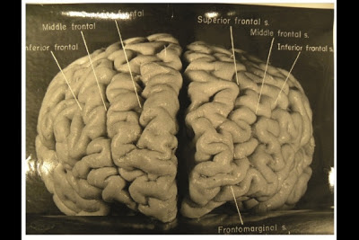
Photographs taken shortly after his death, but never before analysed in detail, have now revealed that Einstein’s brain had several unusual features, providing clues about the neural basis of his extraordinary mental abilities.


Nature.com reports that, while doing Einstein's autopsy, the pathologist Thomas Harvey removed the physicist's brain and preserved it in formalin. He then took dozens of black and white photographs of it before it was cut up into 240 blocks. Now, anthropologist Dean Falk of Florida State University in Tallahassee and her colleagues have obtained 12 of Harvey’s original photographs from the National Museum of Health and Medicine in Silver Spring, Maryland, analysed them and compared the patterns of convoluted ridges and furrows with those of 85 brains described in other studies.Many of the photographs were taken from unusual angles, and show structures that were not visible in photographs that have been analysed previously. The analysis was recently published today in the journal Brain. The most striking observation, says Falk, was “the complexity and pattern of convolutions on certain parts of Einstein's cerebral cortex”, especially in the prefrontal cortex, and also parietal lobes and visual cortex.

The autopsy revealed that Einstein’s brain was smaller than average and subsequent analyses showed all the changes that normally occur with ageing. Nothing more was analysed, however. Harvey stored the brain fragments in a formalin-filled jar in a cider box kept under a beer cooler in his office. Decades later, several researchers asked Harvey for some samples, and noticed some unusual features when analysing them.
A study done in 1985 showed that two parts of his brain contained an unusually large number of non-neuronal cells called glia for every neuron2. And one published more than a decade later showed that the parietal lobe lacks a furrow and a structure called the operculum3. The missing furrow may have enhanced the connections in this region, which is thought to be involved in visuo-spatial functions and mathematical skills.
AFP/Getty Images
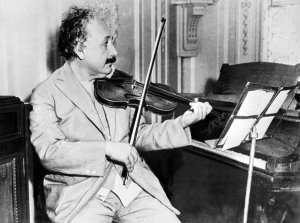
Einstein was a keen violinist, which may account for an overdeveloped section of his brain that deals with the left hand.
The prefrontal cortex is important for the kind of abstract thinking that Einstein would have needed for his famous thought experiments on the nature of space and time, such as imagining riding alongside a beam of light. The unusually complex pattern of convolutions there probably gave the region and unusually large surface area, which may have contributed to his remarkable abilities.
Falk and her colleagues also noticed an unusual feature in the right somatosensory cortex, which receives sensory information from the body. In this part of Einstein’s brain, the region corresponding to the left hand is expanded, and the researchers suggest that this may have contributed to his accomplished violin playing.
According to Sandra Witelson, a behavioural neuroscientist at McMaster University in Hamilton, Canada, who discovered that the parietal operculum is missing from Einstein’s brain, the study’s biggest contribution may be in encouraging further studies. “It makes clear the location and accessibility of photographs and slides of Einstein's brain,” she says. “This may serve as an incentive for other investigations of Einstein's brain, and ultimately of any consequences of its anatomical variations.”

Monday, March 12, 2018

Featured Neuropathologist: Michael Punsoni, MD

On occasion, we profile a prominent or rising neuropathologist. In the past, we've featured the likes of Craig HorbinskiRoger McLendon, Jan Leestma, and Karra Jones. Today we feature Michael Punsoni, MD, a 2016 graduate of the Brown University Neuropathology Fellowship Program and now on faculty at the University of Nebraska in Omaha. Dr. Punsoni agreed to engage in a little Q&A:


1. Why did you decide to become a neuropathologist?
I have always had a strong interest in science and medicine, particularly the neurosciences. After college I worked in two research labs, which fueled my interest in basic neuroscience but also drove me to pursue a medical degree. During my clinical years of medical school I had a strong interest in neurology but my eagerness to be involved in all facets of medical care led me to apply for a categorical residency in Internal Medicine. While I am grateful for the skills and knowledge I acquired during my medicine internship I came to the realization (on one of my 36-hour calls if I remember correctly) that clinical medicine was not for me. I went back to the specialty drawing board and ultimately found pathology somewhat by chance. One of my patients on the medical floor needed an aspiration biopsy of a neck mass. I met the cytopathologist and watched closely as she aspirated a small amount of material and looked on in awe at the squamous cell carcinoma cells on her bedside dual-head scope. I fell for pathology hard after that and, while re-applying to the match, I went back to what I knew best, another year of neuroscience research. By the time I was in pathology residency, my interest for neuropathology was cemented and there was no going back.


2. What do you like to do outside of work?
Watching old and new movies and finding great hole-in-the-wall type restaurants. I’m always looking for/open to suggestions for either one.


3.
 Name a couple of important professional mentors. Why were they important to you?
My two PIs at Cornell Medical Center, Joe Pierce and Theresa Milner for the brilliant work they let me participate in, for teaching me to be meticulous in all things particularly bench techniques and for their good humor that stays with me today. To all four neuropathologists at Brown University who shaped the neuropathologist I would become and still hope to be one day. Dr. Suzanne de la Monte to whom I am grateful for her relentless push to make me a good presenter and for sharing her invaluable tips on manuscript writing. Dr. Douglas Anthony whose leadership skills and commitment to the scientific method were inspiring then and now. Dr. Ed Stopa who treats all his fellows like family and never stops guiding them. Dr. John Donahue who taught me that a remarkable memory is only part of what makes a good pathologist and also, that “it’s a tough job but someone’s got to do it!”.


4.
 What advice would you give to a pathology resident interested in doing a neuropathology fellowship?
Do an elective at your home institution and/or elsewhere. Try it out. It’s a fascinating field and will be for years to come. As I once heard one of my mentors say, we have our own language (when describing the structures of the brain) and we like it that way. Join the group, we’d love to have you.


5. What city (other than Omaha, of course) would you like a future American Association of Neuropathologists meeting to be held and why? 
Honolulu. I’ve never been and this would be a great reason to go. Another desirable place would be Boston, which has great restaurants and a good transit system.

Thursday, March 8, 2018

Cerebral malaria in a young woman who had traveled to Africa

A young woman became sick after visiting Zambia. She died a few weeks after onset of headache and flu-like symptoms. The slide is not dirty! The dirty-looking stuff in the picture below is birefringent pigment called hemozoin. This is typical of a falciparum infection. There are ring hemorrhages and Dürck granulomas present as well. Dürck granulomas are accumulations of mononuclear cells, predominantly macrophages, probably related to resorption of ring hemorrhages.


Hemozoin pigment
Dürck granuloma

Wednesday, March 7, 2018

The meticulously extracted nervous system of a 19th-century woman on display at Hahnemann Medical College

Last summer I put up a post about a remarkable whole nervous system dissection that was carried out at the University of Colorado School of Medicine. The inimitable Dr. Mark Cohen recently sent me an article about a similar dissection performed at Hahnemann Medical College in Philadelphia by Dr. Rufus B. Weaver. The dissection, which took place in 1888 over the course of five months, was performed on a 35-year-old woman who had given permission for her body to be used for the furtherance of science.

Dr. Rufus B. Weaver and the nervous system of Harriet Cole

An excerpt from the article appearing in Atlas Obscura:
According to the History of the Homoeopathic Medical College of Pennsylvania, Dr. Weaver told a fellow doctor about Harriet during a trip to the U.K., after his extraction of the nervous system. He didn’t mention the completion of the dissection. The doctor’s response: “It is impossible, there is no such thing in all this United Kingdom, and if it had been possible it would have been done by some one.” Dr. Weaver replied quietly: “So it has, by some one in the States.” 
In an article for Homeopathic World in August 1892, Dr. Alfred Heath was far more generous about Dr. Weaver’s accomplishment. He called it “a marvel of patience and skill in dissection, the likes of which has never been seen before.”

A dissection similar to that of Dr. Weaver's done at the University of Colorado in 2017 by Shannon Curran

Monday, March 5, 2018

Neuropathology Blog is Signing Off

Neuropathology Blog has run its course. It's been a fantastic experience authoring this blog over many years. The blog has been a source...